- Title
-
A comprehensive structural, lectin and immunohistochemical characterization of the zebrafish olfactory system
- Authors
- Villamayor, P.R., Arana, Á.J., Coppel, C., Ortiz-Leal, I., Torres, M.V., Sanchez-Quinteiro, P., Sánchez, L.
- Source
- Full text @ Sci. Rep.
|
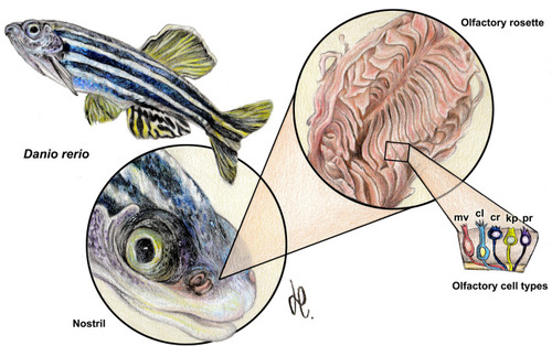
Macroscopic anatomy of the zebrafish olfactory organ. The olfactory rosette occupies in the head an anterodorsal position. The nostrils lie slightly rostral to the eyes and close to the mouth. Five neurosensorial olfactory cell types (OSNs) have been described: microvillous (mv); ciliated (cl); crypt (cr); kappe (kp); and pear (pr) OSNs. Drawing by Helena Reino Piñeiro published under a CC BY open access license. |
|
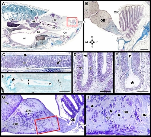
Microscopic anatomy of the zebrafish olfactory system. ( |
|
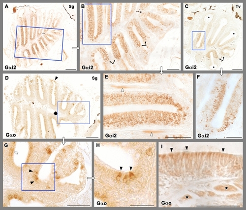
Immunohistochemical study of the olfactory rosette of zebrafish with antibodies against G-proteins. ( |
|
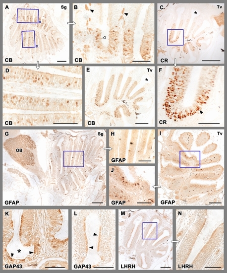
Immunohistochemical study of the olfactory rosette of zebrafish. ( |
|
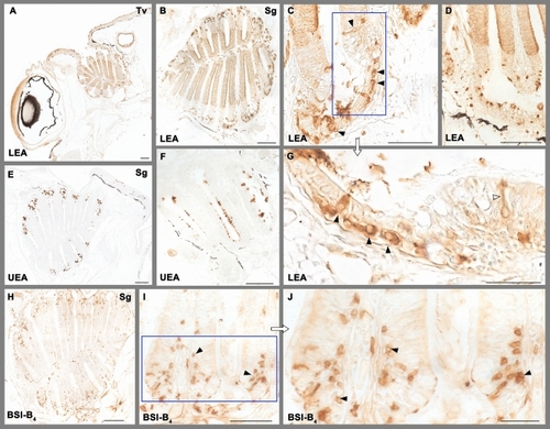
Lectin histochemical staining of the olfactory rosette. ( |
|
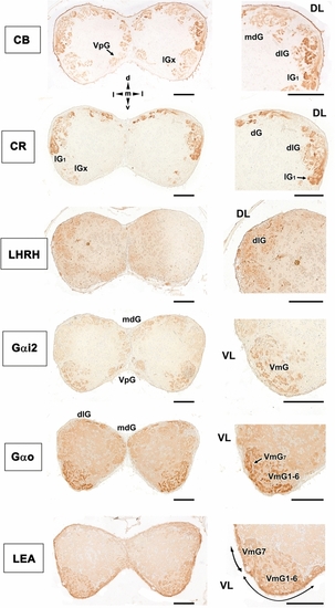
Immunohistochemical and histochemical labelling of the zebrafish olfactory bulb. Transverse sections through the central area of the olfactory bulb showing immunoreactivity with the antibodies anti-CB, anti-CR, anti-LHRH, anti-Gαi2, anti-Gαo and LEA lectin labelling. Calcium-binding proteins (CB and CR) are both mainly expressed in the dorsal (dG), dorsolateral (dlG) and lateral (lGx, lG1) glomeruli. Additionally, calbindin is also expressed in glomeruli belonging to the mediodorsal part of the bulb (mdG) and, with less intensity, in the ventral posterior area (VpG). LHRH is expressed in the dorsolateral glomeruli. Both anti G-proteins show a similar pattern, with an intense labelling in the ventromedial glomerular (VmG) area, stronger in the case of Gαo. Additionally, anti-Gαi2 marks glomeruli belonging to the ventral posterior cluster (VpG). LEA labelling is mainly circumscribed to the ventral region. DL, dorsolateral; VL, ventrolateral; dG, dorsal glomeruli; dlG, dorsolateral glomeruli; lG1, lateral glomerulus 1; lGx, lateral cluster of glomeruli; mdG, mediodorsal glomeruli; VmG1-6, ventromedial glomeruli 1–6; VmG7, ventromedial glomerulus 7; VpG, ventral posterior glomeruli; d, dorsal; l, lateral; m, medial; v, ventral. Scale bars: 100 µm. |

