FIGURE SUMMARY
Title

Functional Characterization of Clinical Isolates of the Opportunistic Fungal Pathogen Aspergillus nidulans

Authors
Bastos, R.W., Valero, C., Silva, L.P., Schoen, T., Drott, M., Brauer, V., Silva-Rocha, R., Lind, A., Steenwyk, J.L., Rokas, A., Rodrigues, F., Resendiz-Sharpe, A., Lagrou, K., Marcet-Houben, M., Gabaldón, T., McDonnell, E., Reid, I., Tsang, A., Oakley, B.R., Loures, F.V., Almeida, F., Huttenlocher, A., Keller, N.P., Ries, L.N.A., Goldman, G.H.
Source
Full text @ mSphere

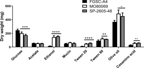
The A. nidulans clinical isolates exhibit improved growth in the presence of alternative carbon and lipid sources. Strains were grown in liquid MM supplemented with glucose, acetate, ethanol, mucin, Tween 20 and 80, olive oil, and Casamino Acids at 37°C for 48 h (glucose) or 72 h (others) before fungal biomass was freeze-dried and weighed. Standard deviations were determined from biological triplicates in a one-way ANOVA with Tukey’s posttest comparing growth of the clinical isolates to that of the FGSC A4 reference strain (*, P < 0.05; **, P < 0.01; ***, P < 0.001; ****, P < 0.0001).

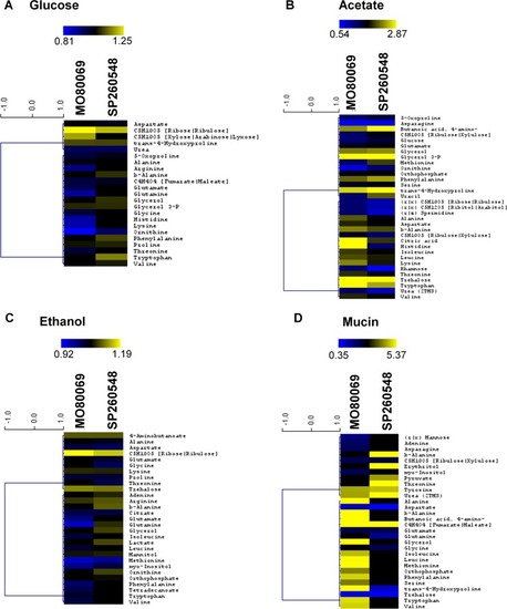
The A. nidulans clinical isolates are metabolically different from the reference strain in the presence of different carbon sources. (A to D) Heat maps depicting log fold changes of identified metabolite quantities that were significantly (P < 0.05) different in the A. nidulans clinical isolates MO80069 and SP-2605-48 compared to levels in the FGSC A4 reference strain (gray squares depict metabolite quantities that were not detected as significantly different in one of the clinical isolates).

The A. nidulans clinical isolates are more sensitive to the cell wall-perturbing agents. (A to C) Strains were grown from 105 spores on glucose minimal medium supplemented with increasing concentrations of caspofungin, Congo red, and calcofluor white for 5 days at 37°C. Standard deviations represent biological triplicates in a two-way ANOVA test, comparing growth of the clinical isolates to growth of the FGSC A4 reference strain (*, P < 0.05; **, P < 0.01; ***, P < 0.001; ****, P < 0.0001).

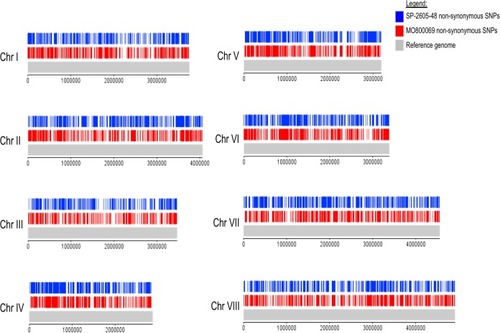
Diagram depicting the location of all detected nonsynonymous single nucleotide polymorphisms (SNPs) on the 8 chromosomes (Chr I to Chr VIII) of the A. nidulans clinical isolates SP-2605-48 and MO80069 in comparison to the FGSC A4 reference genome.

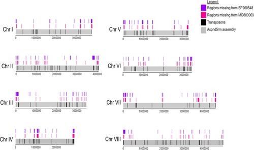
Diagram depicting the location of all detected small deletions on the 8 chromosomes (Chr I to Chr VIII) of the A. nidulans clinical isolates SP-2605-48 and MO80069 in comparison to the FGSC A4 reference genome. Also shown are the locations of putative transposons in the A. nidulans reference genome.

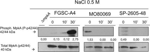
MpkA is not phosphorylated in the A. nidulans clinical isolates MO80069 and SP-2605-48 in the presence of NaCl-induced cell wall stress in contrast to MpkA levels in the FGSC A4 reference strain. Strains were grown from 1 × 107 spores in complete medium for 16 h (control, 0 min) at 37°C before 0.5 M NaCl was added for 10 min (10′) and 30 min (30′). Total cellular protein was extracted, and Western blotting was carried out probing for phosphorylated MpkA. Signals were normalized by the amount of total MpkA present in the protein extracts, and cellular extracts from the ΔmpkA strain were used as a negative control.

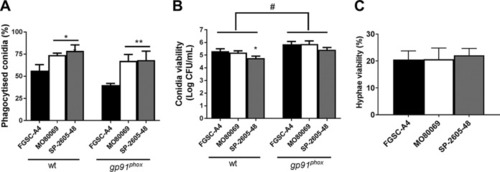
The A. nidulans clinical isolates MO80069 and SP-2605-48 do not present increased survival in the presence of macrophages and neutrophils. (A) Percentage of phagocytized conidia by murine wild-type and gp91phox knockout macrophages. Macrophages were incubated for 1.5 h with conidia from the respective strains before phagocytized conidia were counted. (B) CFU counts as a measure of conidium viability after passage through wild-type (wt) and gp91phox knockout macrophages. Macrophages were incubated with the respective conidia for 1.5 h before they were lysed, and contents were plated on complete medium. (C) Percentage of viable hyphal germlings after incubation for 16 h with neutrophils from healthy human donors. Strain viability was calculated relative to incubation without PMN cells, which was set at 100% for each sample. Standard deviations represent biological triplicates in a one-way ANOVA test with Tukey’s posttest (*, P < 0.05; **, P < 0.01, for results for the clinical isolates compared to those for FGSC A4; #, P < 0.05, for a comparison of results for the two types of macrophages in the same strain).

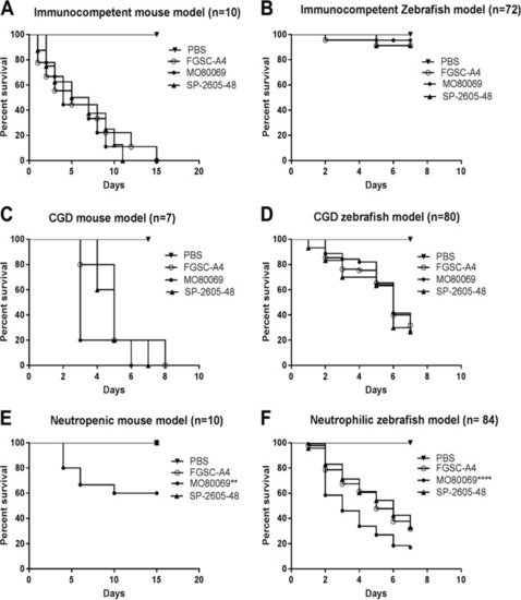
A. nidulans strain-specific virulence depends on the host immune status. The virulence of the A. nidulans clinical isolates MO80069 and SP-260548 was tested in murine (A, C, and E) and zebrafish (B, D, and F) models of pulmonary and invasive aspergillosis. Animals were manipulated in order to give rise to either immunocompetent (A and B), CGD (chronic granulomatous disease) (C and D), or neutropenic (E)/neutrophilic (F) models. Shown are survival curves for each immunosuppression condition and animal model. No difference in virulence levels was detected for all strains in both immunocompetent and CGD mice. Strain MO80069 was significantly more virulent in neutropenic mice and neutrophilic zebrafish. **, P < 0.01; ****, P < 0.0001 for a comparison of the values for the clinical isolates to those of the FGSC A4 reference strain in a two-way ANOVA test with Tukey’s posttest.

Acknowledgments
This image is the copyrighted work of the attributed author or publisher, and ZFIN has permission only to display this image to its users. Additional permissions should be obtained from the applicable author or publisher of the image. Full text @ mSphere