FIGURE SUMMARY
Title

Intracellular Localization in Zebrafish Muscle and Conserved Sequence Features Suggest Roles for Gelatinase A Moonlighting in Sarcomere Maintenance

Authors
Fallata, A.M., Wyatt, R.A., Levesque, J.M., Dufour, A., Overall, C.M., Crawford, B.D.
Source
Full text @ Biomedicines

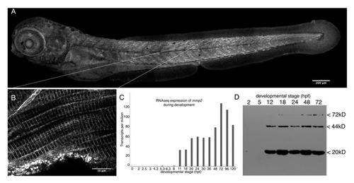
Mmp2 is expressed ubiquitously from early development and accumulates in a striated pattern within the skeletal muscle. (A) Composite confocal projections of a 72 hpf embryo stained with anti-Mmp2 exhibiting labeling throughout the embryo with notable accumulation in the skeletal muscle (scale bar = 200 µm). (B) High magnification view of a single confocal section through the trunk musculature (indicated by the inset) showing strong labeling of the myotome boundary (upper left corner), and striated staining in myofibrils (scale bar = 10 µm). (C) RNASeq data showing absolute abundance of mmp2 transcripts in embryos from fertilization to five days post-fertilization (dpf). (D) Immunoblot of whole embryo homogenates (350 µg per lane) made from 2 hpf (cleavage), 5 hpf (50% epiboly), 12 hpf (early somitogenesis), 18 hpf (late somitogenesis), 24 hpf (prim-5), 48 hpf (long pec), and 72 hpf (protruding mouth) embryos probed with anti-Mmp2. Immunoreactivity is detected in embryos after 12 h of development, with bands at the expected mobility for full-length Mmp2 (72 kD) and stronger bands at 44 and 20 kD, which combine to give the expected size for activated Mmp2 (64 kD).

EXPRESSION / LABELING:
Gene:
Antibody:
Fish:
Anatomical Term:
Stage: Protruding-mouth

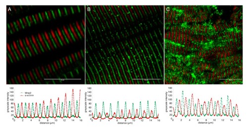
Mmp2 is localized between Z-discs in sarcomeres of embryonic and adult muscle. Confocal micrographs of skeletal muscle from 72 hpf embryos (A) or adults (B), and 500 nm thick cryosection of 72 hpf skeletal muscle (C) stained with anti-α-actinin (red) and anti-Mmp2 (green). Greyscale intensity profiles of both channels along a line drawn perpendicular to the sarcomeres are shown below each micrograph. Mmp2 immunoreactivity occurs at regularly spaced intervals precisely between the α-actinin-labeled Z-discs. Scale bars = 10 µm.

EXPRESSION / LABELING:
Gene:
Antibody:
Fish:
Anatomical Term:
Stage: Protruding-mouth

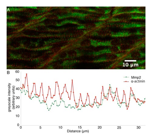
Sarcomeric Mmp2 begins to accumulate subsequent to the assembly of Z-disks. (A) Confocal section through the trunk musculature of a 24 hpf embryo posterior to the yolk extension, at the position at which myofibrils are differentiating. (B) Sarcomeric α-actinin (red) is beginning to become apparent as Z-bodies in the periphery of differentiating myocytes, but Mmp2 immunoreactivity (green) remains roughly homogeneously distributed. Greyscale intensity profiles of both channels along a line drawn through the periphery of a differentiating myocyte are shown below the micrograph. Scale bar = 10 µm.

EXPRESSION / LABELING:
Gene:
Antibody:
Fish:
Anatomical Term:
Stage: Prim-5

The secretory signal peptide of gelatinase A orthologues is consistently and significantly less likely to be recognized than that of most other type-I secreted proteins. Violin plots of mean ‘S’ scores of the N-terminal secretory signals from orthologues of vitronectin (Vtn) and all secreted MMPs. MMPs with mean S score statistically indistinguishable from vitronectin are shown in blue. Mean S scores for orthologues of gelatinase A (red) are significantly lower than those of vitronectin and other secreted MMPs apart from MMP21 and MMP23, which undergoes type II secretion and therefore, does not have an N-terminal secretory signal. Statistically, indistinguishable groups are indicated with letters at the top of the plot, and the number of orthologues of each protein analyzed is indicated along the x-axis.

Gelatinase A orthologues have highly conserved phosphorylation sites. Putative serine (solid lines), threonine (dashed lines), and tyrosine (dotted lines) phosphorylation sites conserved in 100% (black), 99% (dark grey), or 97% (light grey) of gelatinase A orthologues are shown with respect to a structural schematic of the gelatinase A protein, illustrating the signal sequence (1–29 (orange)), propeptide (30–107 (grey)), catalytic domain (118–446 (green)) with fibronectin-like repeats (light green), and hemopexin-like domain (463–657 (purple)). Cysteines are indicated with yellow spots, connected by horizontal lines if they are predicted to participate in intramolecular disulfide bonds. Conserved residues that have been empirically demonstrated to be phosphorylated in vivo in the human protein are indicated with asterisks.

Acknowledgments
This image is the copyrighted work of the attributed author or publisher, and ZFIN has permission only to display this image to its users. Additional permissions should be obtained from the applicable author or publisher of the image. Full text @ Biomedicines