FIGURE SUMMARY
Title

Reactivation of Notch signaling is required for cardiac valve regeneration

Authors
Kefalos, P., Agalou, A., Kawakami, K., Beis, D.
Source
Full text @ Sci. Rep.

Chemically induced genetic cell ablation of zebrafish larval cardiac valves. (A) Outline of chemical treatment of UAS-E1b:NfsB-mCherry transgenic embryos with Metronidazole (MTZ). Metronidazole was applied at 3.5 dpf embryos and washed-off at 4 dpf. Embryos were then left for recovery in order to observe regeneration process. (B,C) Gfp and mCherry Gal4 driven expression in 4 dpf cardiac valves (Arrows). AV: atrioventricular canal, (BA): bulbus arteriosus. Scale bars: 100 μm. (D) Confocal z stack projection of a double transgenic Tg(hspGFFDMC73A/UAS-E1b:NfsB-mCherry) embryonic heart stained with cell-cell adhesion marker Dm:grasp (blue). Scale bar: 20 μm (F,G) Gfp and mCherry Gal4 driven expression in 4 dpf cardiac valves of MTZ treated embryos. Injury is observed at AV canal and BA nfsb-mCherry+ cells that have been ablated. Scale bars: 100 μm. (H) Confocal z-stack projection of a double transgenic Tg(hspGFFDMC73A/UAS-E1b:NfsB-mCherry) heart treated with MTZ and stained with cell-cell adhesion marker Dm-grasp (blue). Arrowheads depict the position where AV canal and BA differentiated cells should be. Scale bar: 20 μm. (E–I) Snapshots of high frame live videos of a DMSO treated and a MTZ-treated Tg(gSAIzGFFD703A/UAS-E1b:NfsB-mCherry) embryo 4 dpf, respectively (S. Movies 3 and 4). Bars show the haemodynamic flow patterns at the atriovantricular canal of uninjured and injured valves per heartbeat. The number of frames per total frames of a heartbeat was measured for forward flow (+) no flow (0) and reverse flow (−). The reverse flow fraction is increased 4,91 times (quantified from n = 8 ctrl embryos and 8 MTZ-treated embryos. p < 0.001 using paired t-test) upon valve ablation. +: forward fraction. 0: null fraction. −: reverse fraction.

Zebrafish embryonic valves can regenerate. (A–C) Confocal z-stacks of untreated Tg(hspGFFDMC73A/UAS-E1b:NfsB-mCherry) larvae at 4 dpf, 6 dpf and 12 dpf respectively. (D–F) Confocal z-stacks of MTZ treated Tg(hspGFFDMC73A/UAS-E1b:NfsB-mCherry) and larvae at 4 dpf, 6 dpf and 12 dpf respectively. Arrows show the ablation of mCherry+ cells at the AV canal and OFT at 4 dpf. From 6 dpf more mCherry+ cells could be observed at the AV canal. By 12 dpf the regeneration process could be imaged, as mCherry+, AV differentiated cells were present at both valves. Scale bars: 20 microns. Each experiment was carried out at least 3 independent times with n = 10 embryos per experimental group.

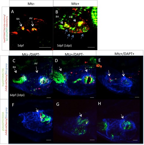
Notch signaling is activated during valve regeneration. (A) Confocal z-stack of a Tg(TP1:VenusPEST)/hspGFFDMC73A: UAS-E1b:NfsB-mCherry double transgenic embryo at 5 dpf. Tg(TP1:VenusPEST) signal of activated Notch signaling is restricted to the valves. Scale bar: 100 microns. (B) 1 day post MTZ treatments. Expansion of activation of Notch signaling throughout the ventricle is observed (blue arrows) Scale bar: 50microns. Confocal z-stacks of untreated Tg(hspGFFDMC73A: UAS-E1b:NfsB-mCherry) (C) and Tg(GSAIzGFFD703A: UAS-E1b:NfsB-mCherry) (F) double transgenic embryos at 6 dpf. Confocal z-stack of MTZ treated Tg(hspGFFDMC73A: UAS-E1b:NfsB-mCherry) (D), and Tg(GSAIzGFFD703A: UAS-E1b:NfsB-mCherry) (G), at 6 dpf. Confocal z-stack of MTZ treated and then during regeneration 48 hours DAPT treated Tg(hspGFFDMC73A: UAS-E1b:NfsB-mCherry) (E), and Tg(GSAIzGFFD703A: UAS-E1b:NfsB-mCherry) (H), at 6 dpf. (I) Each experiment was carried out at least 3 independent times with n = 10 embryos per experimental group. Scale bars: 20 micron.

Zebrafish adult valves retain the ability to regenerate through Notch signaling reactivation. (A–D) Z-stack projection of a wt untreated adult valve of an hspGFFDMC73A/UAS-E1b:NfsB-mCherry transgenic fish stained with elastin2. (E–H) Valvular damage of an adult heart of a hspGFFDMC73A/UAS-E1b:NfsB-mCherry transgenic immediately after completion of the MTZ treatment (n = 10). (I–L) Regenerating valve 15 days post chemogenetic valve cell ablation (n = 13). (M–P) Inhibition of regeneration process in the heart of an adult heart of a hspGFFDMC73A/UAS-E1b:NfsB-mCherry transgenic after MTZ treatment and 15 day treatment with DAPT (n = 12). Scale bars: 50 micron.

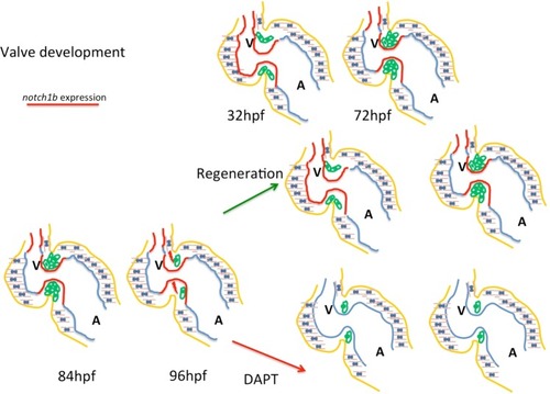
Sensing of immature intracardiac flow patterns activates a Notch mediated regenerative program, recapitulating cardiac valve development. At embryonic developmental stages a feedback loop where intracardiac flow dynamics regulate klf2a and notch1b results in the restricted expression of notch1b at the high sheer-stress areas of the heart: the atrioventricular valve and the outflow tract. The intracardiac flow pattern following valvular damage, is disturbed causing an increase in the retrograde blood flow fraction, reminiscent of the flow pattern of earlier stage of embryogenesis. As a result, notch1b is re-activated ectopically the ventricular endocardium. When cardiac valves regenerate and flow dynamics are restored, notch1b expression gets restricted at the atrioventricular canal and the outflow tract.

Acknowledgments
This image is the copyrighted work of the attributed author or publisher, and ZFIN has permission only to display this image to its users. Additional permissions should be obtained from the applicable author or publisher of the image. Full text @ Sci. Rep.