- Title
-
Notch-mediated inhibition of neurogenesis is required for zebrafish spinal cord morphogenesis
- Authors
- Sharma, P., Saraswathy, V.M., Xiang, L., Fürthauer, M.
- Source
- Full text @ Sci. Rep.
|
Mindbomb1 is required for zebrafish spinal cord morphogenesis. (a,b) Morpholino knock-down of mib1 disrupts apical aPKC enrichment and neuro-epithelial morphology (outlined by F-actin staining) in 15/19 embryos. (c) A similar disruption of apico-basal polarity is observed in mib1 mutants (n = 14). (d,e) mib1 mutants fail to establish polarized Pard3 localization (e, n = 12). (f,g) No polarized Crumbs enrichment is observed in mib1 mutants (g, n = 17). (h,i) The apical localization of the tight junction component ZO-1 is disrupted in mib1 mutants (i, n = 16). (j,k) Centrosomes fail to move towards the neural tube midline in mib1 mutants (k, n = 16). (a–c,f,g) 30 somites stage. d,e, 18 somites stage, (h–k) 22 somites stage. All images are dorsal views of the anterior spinal cord, anterior left. Scalebars: 20 µm.
|
|
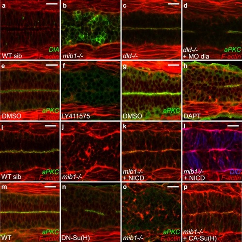
Notch pathway activity is required for spinal cord morphogenesis. ( |
|
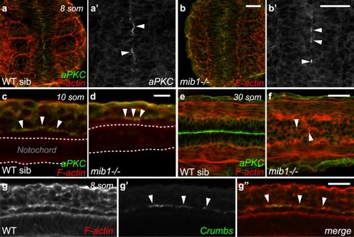
Notch signaling is dispensable for the emergence of floor plate apico-basal polarity. ( |
|
Notch signaling is required for the establishment of apico-basal polarity in the dorso-medial spinal cord. ( |
|
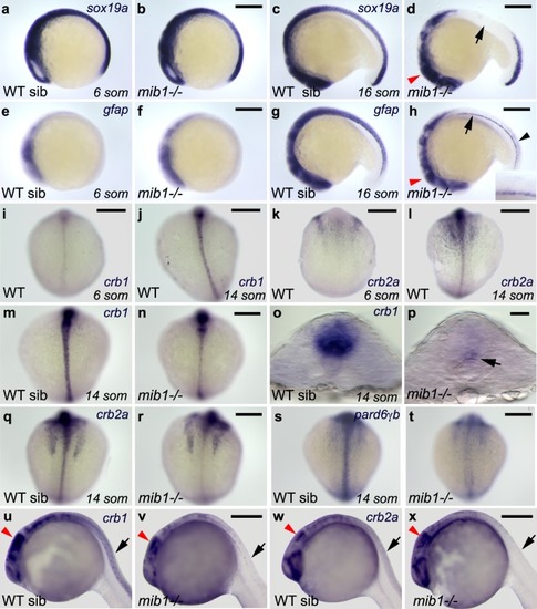
Notch signaling is required to allow the emergence of neuroepithelial identity. (a,b) The expression of the neuronal progenitor marker sox19a is similarly initiated in WT sibling (a) and mib1 mutant embryos (b, n = 4). (c) By the 16 somites stage, sox19aexpression is still present in the brain and anterior spinal cord of WT siblings. (d) In mib1 mutants (n = 17) sox19a is lost in the anterior spinal cord (arrow) but partially retained in the brain (red arrowhead). (e,f) At the 6 somites stage, low levels of the radial glia marker gfap are detected in the brain region of WT siblings (e) or mib1mutants (f, n = 10). (g) By the 16 somites stage, WT siblings display gfap expression in the brain and spinal cord. (h) mib1 mutants (n = 17) fail to upregulate gfapexpression in the dorso-medial anterior spinal cord (inset). Reduced gfap expression levels are observed in the brain (red arrowhead) and caudal spinal cord (black arrowhead). Continuous gfap expression is retained in the floor plate (arrow). (i–l) The establishment of apico-basal polarity coincides with an upregulation of crb1 (i,j) and crb2a (k,l) in the spinal cord. (m,n) Upregulation of crb1 expression is impaired in mib1 mutants (n, n = 11). (o,p) Residual crb1 expression persists in the ventral-most spinal cord (arrow in p) of 14 somites stage mib1 mutants. (q,r) mib1 mutants display reduced crb2a expression in the spinal cord (r, n = 13). (s,t) pard6γb expression is lost in the neural tube of mib1 mutants (t, n = 13). (u–w) In 30 somites stage mib1mutants crb1 (b, n = 10) and crb2a (d, n = 10) expression are lost in the anterior spinal cord (black arrows) but partially retained in the brain (red arrowheads indicate the midbrain). (a–h, u–x) lateral views, anterior to the left, dorsal up. (i–n, q–t) dorsal views of the spinal cord, anterior up. (o,p) transversal sections of the neural tube, dorsal up. Scalebars: (a–n,q–t) 200 µm, (o,p) 20 µm, (u–x) 250 µm.
|
|
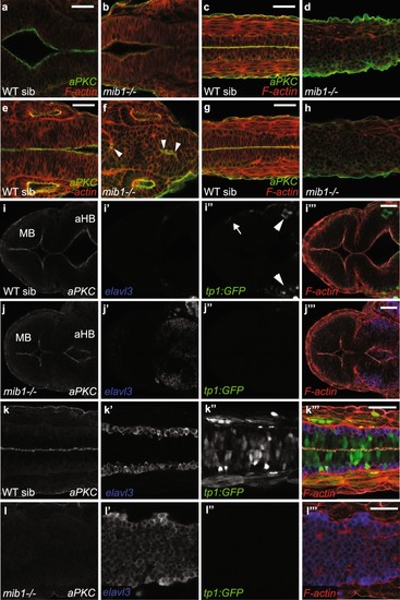
Notch loss of function differentially affects neuroepithelial polarity in the brain and spinal cord. ( |
|
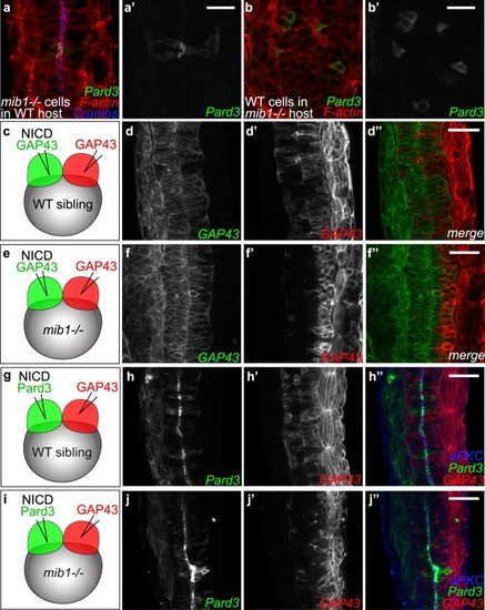
Dissection of the spatial requirement for Notch signaling in neural tube morphogenesis. ( |
|
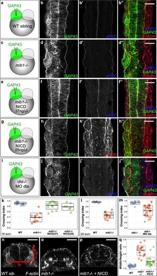
Notch loss of function impairs morphogenetic cell movements in the zebrafish spinal cord. ( |


