- Title
-
Development of zebrafish medulloblastoma-like PNET model by TALEN-mediated somatic gene inactivation
- Authors
- Shim, J., Choi, J.H., Park, M.H., Kim, H., Kim, J.H., Kim, S.Y., Hong, D., Kim, S., Lee, J.E., Kim, C.H., Lee, J.S., Bae, Y.K.
- Source
- Full text @ Oncotarget
|
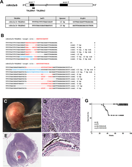
Somatic inactivation of cdkn2a/b gene by the injection of TALENs mRNA leads to MPNSTs in F0 founder zebrafish. (A) The targeted regions of two different TALENs (cdkn2a/b TALENs-1 and cdkn2a/b TALENs-2) are located in exon 1 of cdkn2a/b gene. The binding sequences of TALENs for cdkn2a/b gene are also shown. (B) By the injection of cdkn2a/b TALENs-1 mRNA, 7 different germline mutated alleles of cdkn2a/b gene were recovered at F1 generation. And 9 different alleles were also recovered at F1 generation after injection of cdkn2a/b TALENs-2 mRNA. (C) The large mass of tumor was observed in adjacent to eyes of the cdkn2a/b TALENs mRNA injected tp53 mutant zebrafish at 5.5 month post fertilization. (D?F) Histological image of cross sections of tumor tissue was visualized with Hematoxylin-Eosin staining. (D) High magnification image of tumor tissue shows the herringbone pattern that is a typical feature of malignant peripheral nerve sheath tumor. (F) There is no evident abnormality in the neuroepithelial layer of retina adjacent to tumor tissue. (G) Kaplan-Meier survival representation of wild type (AB), tp53 mutant, and cdkn2a/b TALENs mRNA injected AB or tp53 mutant zebrafish. The cdkn2a/b TALENs mRNA injected tp53 mutant zebrafish had only started to exhibit death with tumor bearing as early as about 5 months. Other adult zebrafish such as tp53 mutant and cdkn2a/b TALENs mRNA injected AB zebrafish did not show early death with tumor bearing. All data was acquired from two independent experiments. Scale bars: 2 mm (E) and 100 ?m (D and F). PHENOTYPE:
|
|
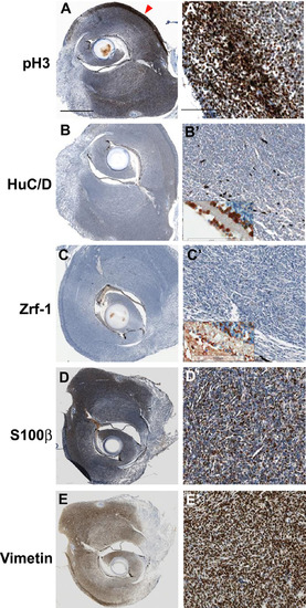
Immunohistochemical analysis of tumors from cdkn2a/b TALENs mRNA injected tp53 mutant zebrafish. (A) (A') Immunostaining with anti- phospho-Histone 3 antibidy. The tumor tissue from cdkn2a/b TALENs mRNA injected tp53 mutant zebrafish is highly mitotic. The phospho-Histone 3 positive mitotic cells were more evident in the outer region of tumor tissue (red arrowhead in A). (B) (B') Neuronal signature that was visualized by immunostaining with anti-HuC/D antibody was scarcely detected in densely cellular region of tumor tissue. (C) (C') Immunostaining with zrf-1 which detect zebrafish GFAP protein. The tumor tissue induced with cdkn2a/b TALENs mRNA also did not show any glial characteristics. Normal staining of anti-HuC/D and zrf-1 antibody could be observed in retinal layers near to tumor tissues (Inset box in B' and C'). (D) (D') Epithelioid MPNST marker, S100?, is strongly expressed in tumor tissue. (E) (E') Mesenchymal marker, Vimentin, is also positively stained in tumor tissue. Scale bars: 1 mm (A) and 100 ?m (A'). EXPRESSION / LABELING:
|
|
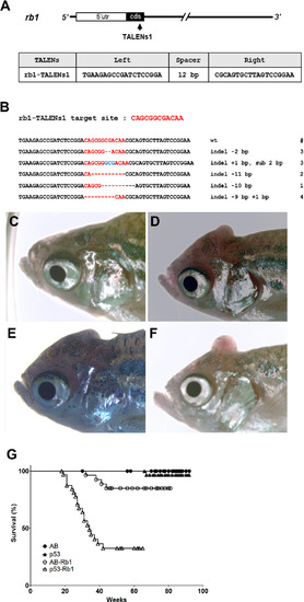
Somatic inactivation of rb1 by the injection of rb1-TALENs mRNA leads to medulloblastoma like PNETs in F0 founder zebrafish. (A) The targeted region of rb1-TALENs is located in 1st exon of zebrafish rb1 locus. The binding sequences of TALENs for rb1 are indicated. (B) After the injection of rb1-TALENs mRNA, 4 different deletion and 1 insertion germline mutated alleles of rb1 were successfully recovered at F1 generation. Hence, 13 independent lines which have frameshift mutation in rb1 could be obtained. (C?F) Morphological appearance of tumor bearing in head region of 5 month old zebrafish was presented. Protruding tumor mass were observed in rb1-TALENs mRNA injected zebrafish. The same aged wild type (C) and mutant (D?F) zebrafish are shown for comparison. (G) Kaplan-Meier survival representation of wild type, tp53 mutant, and rb1-TALENs injected wild type or tp53 mutant zebrafish. At about 6 month post fertilization, zebrafish which were injected with rb1-TALENs mRNA at one cell stage had started to die with tumors in the head region in wild type adult zebrafish. The tumor incidence which was induced by the injection of rb1-TALENs mRNA was more evident in tp53 mutant zebrafish. Adult zebrafish that showed abnormal swimming behavior started to die with tumor in head regions as early as about 20 weeks after injection of rb1-TALENs mRNA into one cell stage embryos. PHENOTYPE:
|
|
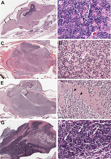
Histopathology of tumors from rb1-TALENs injected tp53 mutant zebrafish. (A) Sagittal section image of H & E staining of wild type zebrafish at 5 month post fertilization. Black arrows and arrow heads indicate forebrain and optic tectum, respectively. Cerebellum and cerebellar granule cells are indicated by white arrows and arrow heads, respectively. (B) High magnitude image of A. Normal cerebellar granule cells have a unique feature that is characterized with small rounded nuclei. (C?H) Sagittal section images of H & E staining of tumors from rb1-TALENs injected tp53 mutant zebrafish. (C) Tumors were mainly arising in cerebellum, medulla, and brainstem. (D) High magnitude image of C. Highly cellular tumor cells with wedged nuclei were observed. Rosette like structures was marked by white arrows. (E) Tumors were mainly arising in cerebellum encompassed a majority of medulla, hypothalamus, and brainstem. The forth ventricle which were surrounded with tumor cells were marked by black arrows. (F) High magnitude image of E. Infiltration of tumor cells was indicated by black arrowheads in dorsal brainstem. (H) High magnitude image of G. Homer-Wright rosettes which are seen in PNETs or medulloblastomas were observed distinctly (white arrows). Anterior is left of all images. Scale bars: 500 ?m (A, C, E and G), and 50 ?m (B, D, F and H). PHENOTYPE:
|
|
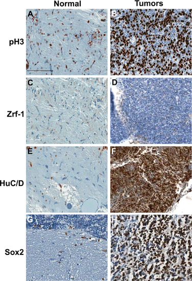
Immunohistochemical analysis of tumors from rb1-TALENs injected tp53 mutant zebrafish. Immunostaining was performed with lineage specific antibody with normal brain tissues (A, C, E, and G) and tumors from rb1-TALENs injected tp53 mutant zebrafish (B, D, F, and H) using anti phospho-Histone 3 (A and B), anti Zrf-1 (C and D), anti HuC/D (E and F) and anti Sox2 (G and H) antibodies. Highly mitotic tumor cells could be detected in tumor tissues from rb1-TALENs mRNA injected tp53 mutant zebrafish (B). Glial marker, Zrf-1 was scarcely stained in densely cellular region of tumor tissue (D). However, HuC/D antigen which is a postmitotic neuronal marker was strongly expressed in densely cellular region of tumor tissue (F). Neural progenitor marker, Sox2 expression was also detected in same cellular region of tumor tissue (H). Scale bars: 50 ?m. EXPRESSION / LABELING:
|
|
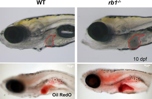
TALEN-mediated mutations of rb1 resulted in a marked enlarged liver in zebrafish larvae. Homozygous rb1 mutant larvae were generated by incrossing heterozygous F1 zebrafish with same frameshift mutated alleles (11bp deletion). rb1 mutants exhibited flattened swimming bladder, and enlarged liver at 7 day post fertilization (dpf) and did not survive until 10 dpf (red broken line). The increased lipid accumulation in liver of rb1 mutants was visualized by Oil Red O staining. WT: wild type embryo, rb1?/?: homozygous rb1 mutant. PHENOTYPE:
|

