
|
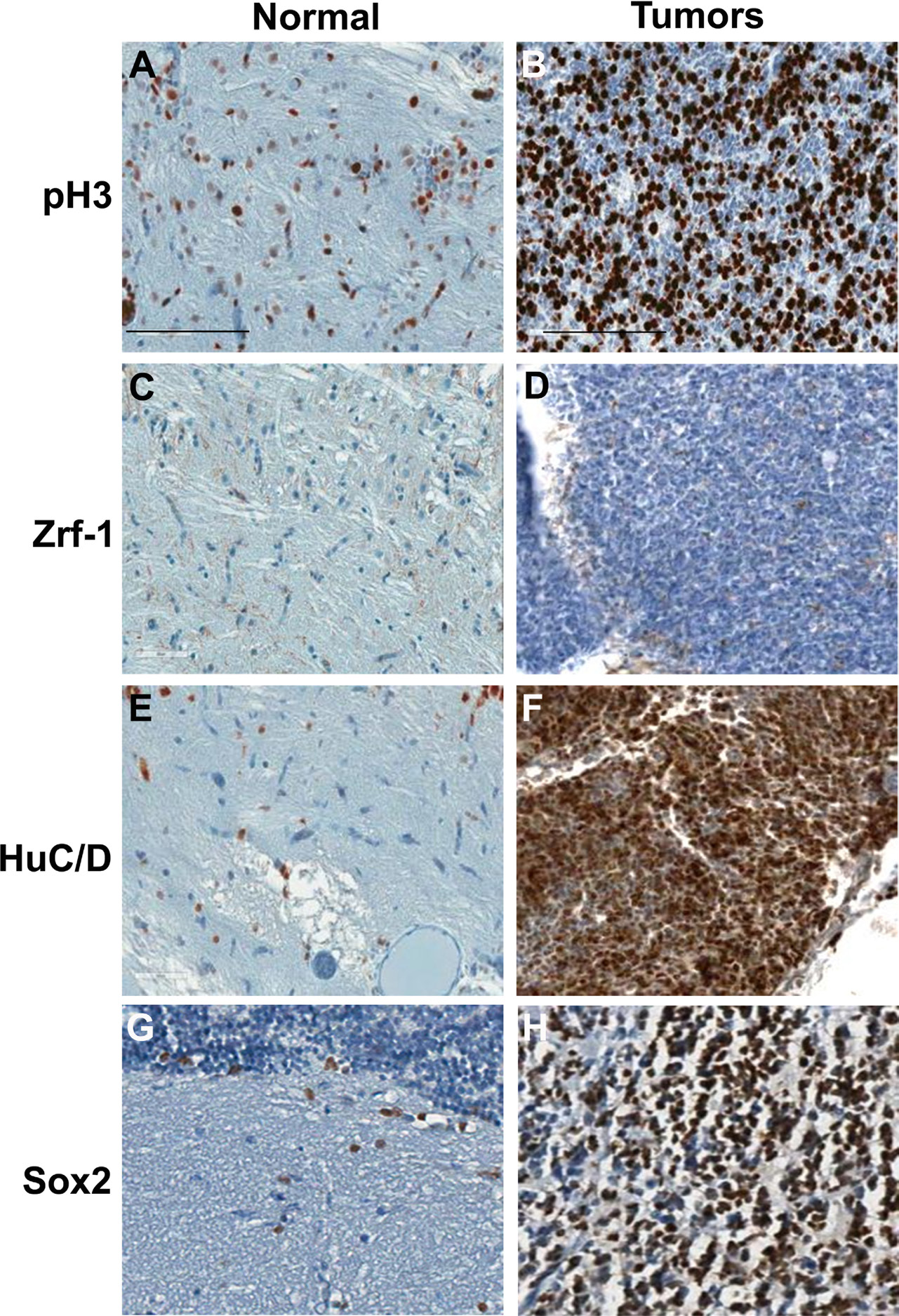
Fig. 5
Immunohistochemical analysis of tumors from rb1-TALENs injected tp53 mutant zebrafish. Immunostaining was performed with lineage specific antibody with normal brain tissues (A, C, E, and G) and tumors from rb1-TALENs injected tp53 mutant zebrafish (B, D, F, and H) using anti phospho-Histone 3 (A and B), anti Zrf-1 (C and D), anti HuC/D (E and F) and anti Sox2 (G and H) antibodies. Highly mitotic tumor cells could be detected in tumor tissues from rb1-TALENs mRNA injected tp53 mutant zebrafish (B). Glial marker, Zrf-1 was scarcely stained in densely cellular region of tumor tissue (D). However, HuC/D antigen which is a postmitotic neuronal marker was strongly expressed in densely cellular region of tumor tissue (F). Neural progenitor marker, Sox2 expression was also detected in same cellular region of tumor tissue (H). Scale bars: 50 ?m.