- Title
-
Analysis of Zebrafish Kidney Development with Time-lapse Imaging Using a Dissecting Microscope Equipped for Optical Sectioning
- Authors
- Perner, B., Schnerwitzki, D., Graf, M., Englert, C.
- Source
- Full text @ J. Vis. Exp.
|
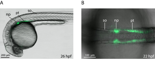
Transgenic wt1b:GFP Embryos with Green Fluorescence in the Developing Kidney. Overlays of dorsal (A) or lateral (B) transmission and fluorescence images are shown with anterior to the left. np, nephron primordium; pt, pronephric tubule; so, somite. EXPRESSION / LABELING:
|
|
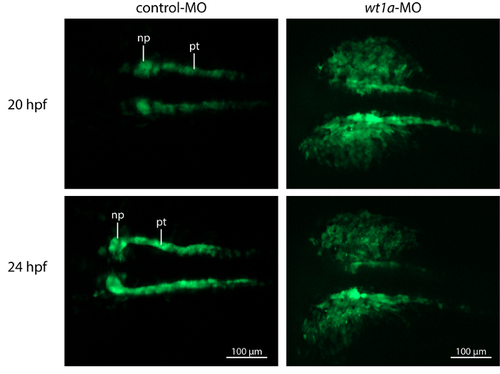
Knockdown of wt1a Disrupts Embryonic Kidney Development. Representative, extended depth of focus images from time-lapse recordings. In control morpholino injected embryos, kidney development shows normal progress with growing tubules and nephron primordia which start to fuse at the midline. In contrast, wt1a morphants fail to form proper nephron primordia and a massive amount of GFP positive cells are outside of the pronephric field. (np, nephron primordium; pt, pronephric tubule). EXPRESSION / LABELING:
PHENOTYPE:
|


