- Title
-
Paxillin and Focal Adhesion Kinase (FAK) Regulate Cardiac Contractility in the Zebrafish Heart
- Authors
- Hirth, S., Bühler, A., Bührdel, J.B., Rudeck, S., Dahme, T., Rottbauer, W., Just, S.
- Source
- Full text @ PLoS One
|
Targeted knock-down of Paxillin leads to heart failure in zebrafish. (A-C) Co-immunostaining of Paxillin (A, red) and the known Z-disk protein Focal Adhesion Kinase (FAK) (B, green) on adult primary zebrafish cardiomyocytes revealed localization of Paxillin to sarcomeric Z-disks. Cell nuclei were counterstained with DAPI (C, blue). Scale bar 10 ?m. (D-G) Lateral view of paxillin start MO (MO1-paxillin) (D) and paxillin 5bp-mismatch-MO (MO1-control) (E) injected embryos at 72 hpf. (F) 78% of embryos injected with 5.4 ng MO1-paxillin developed pericardial edema and blood congestion at the cardiac inflow tract (n = 3; *P<0.0001), whereas control-injected embryos developed no pathological phenotype. (G) Fractional shortening (FS) measurements of Paxillin morphant ventricles at 48, 72 and 96 hpf. FS of Paxillin morphant ventricles was slightly reduced to 69.6% ± 5.07% compared to corresponding 5bp-mismatch-MO injected embryos (FS: 71.6% ± 3.96%) at 48 hpf. At 72 hpf, FS in Paxillin morphants was reduced to 49.29% ± 4.39% compared to control morphants (FS: 69.3% ± 4.36%), whereas ventricular chambers of Paxillin morphants became almost silent (FS: 4.23% ± 6.17%) compared to controls (FS: 74% ± 4.96%) at 96 hpf (n = 6?8 individuals per time point). (H-K) Lateral view of MO2-paxillin (H) and MO2-paxillin+paxillin mRNA (I) injected embryos at 72 hpf. (J) Co-injection of 4.5 ng of MO2-paxillin and 1 ng wild-type paxillin mRNA rescued the Paxillin morphant heart failure phenotype (n = 3; *P = 0.0073). (K) Quantification of ventricular FS revealed that ectopic expression of paxillin mRNA significantly improved contractile function in Paxillin morphants at 72 hpf (n = 8?9 individuals; *P<0.0001). Error bars indicate s.d. EXPRESSION / LABELING:
PHENOTYPE:
|

ZFIN is incorporating published figure images and captions as part of an ongoing project. Figures from some publications have not yet been curated, or are not available for display because of copyright restrictions. EXPRESSION / LABELING:
|
|
Knock-down of fak1a and fak1b phenocopies the Paxillin morphant phenotype. (A) Western blot analysis of FAK protein levels in Paxillin-deficient zebrafish embryos compared to control-injected embryos. For each sample 50 embryos were pooled and 20 ?g of protein lysate were loaded per lane. The figure shows one representative western blot out of three independent experiments. (B, C) Quantitative real time PCR of Paxillin-depleted and control-injected embryos showed no significant (ns) alteration of fak1a (n = 3; *P = 0.9017) (B) and fak1b (n = 3; *P = 0.8878) (C) mRNA expression. For statistical analysis student?s t-test was performed. (D-F) Lateral view of MO1-fak1a/fak1b (D) and 5bp-mismatch-MOs (MO1-control) (E) injected embryos at 72 hpf. Embryos co-injected with 2.7 ng of MO1-fak1a and 2.15 ng of MO1-fak1b showed a severe heart failure phenotype whereas injection of the same amount of fak1a and fak1b 5bp-mismatch MOs (MO1-control) caused no significant pathological cardiac phenotype (F) (n = 3; *P<0.0001). (G) FS measurements of FAK morphant ventricles at 36, 48 and 72 hpf. FS of FAK double-knock-down morphant ventricles was slightly reduced to 56.38% ± 5.85% compared to corresponding 5bp-mismatch-MO injected embryos (FS: 62.4% ± 5.77%) at 36 hpf. At 48 hpf, FS in FAK morphants was reduced to 63.57% ± 9.18% compared to control morphants (70.33% ± 3.72%), whereas ventricular chambers of FAK-deficient embryos became almost silent (FS: 2.6% ± 3.71%) compared to controls (FS: 69.33% ± 4.36%) at 72 hpf (n = 5?8 individuals per time point). (H) Western blot analysis of Paxillin levels in control-, MO1-fak1a/fak1b- and MO1-paxillin-injected embryos. Paxillin protein levels are severely reduced after targeted knock-down of FAK and Paxillin, respectively. For each sample 50 embryos were pooled and 20 ?g of protein lysate were loaded per lane. The figure shows one representative western blot out of three independent experiments. (I) Quantitative real time PCR of FAK-depleted and control-injected embryos showed no significant (ns) alteration of paxillin mRNA expression (n = 3; *P = 0.0937). For statistical analysis student?s t-test was performed. (J) Western blot analysis of embryos co-injected with zebrafish paxillin mRNA and MO2-paxillin compared with embryos injected with MO2-paxillin or paxillin mRNA alone. Ectopic expression of zebrafish paxillin mRNA was able to restore FAK protein expression. For each sample 50 embryos were pooled and 20 ?g of protein lysate were loaded per lane. Error bars indicate s.d. EXPRESSION / LABELING:
PHENOTYPE:
|

ZFIN is incorporating published figure images and captions as part of an ongoing project. Figures from some publications have not yet been curated, or are not available for display because of copyright restrictions. |
|
Vinculin does not localize to focal adhesion sites in Paxillin- and FAK-deficient zebrafish embryos. (A-C) Co-immunostaining of dissected embryonic (72 hpf) zebrafish hearts with antibodies against ?-Catenin (red) and Vinculin (green) of control- (A), MO1-fak1a/fak1b- (B) and MO1-paxillin- (C) injected hearts. Arrow heads indicate focal adhesion sites. Scale bar 5?M. EXPRESSION / LABELING:
PHENOTYPE:
|
|
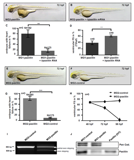
Injection of human paxillin mRNA rescues the cardiac phenotype of MO1-Paxillin morphants. (A-C) Lateral view of MO1-paxillin (A) and MO1-paxillin+human paxillin mRNA (B) injected embryos at 72 hpf. (C) Bar graphs compare average of rescued embryos after co-injection of human paxillin mRNA compared to MO1-paxillin injected embryos (n = 3; *P = 0.0073). (D) Fractional shortening (FS) measurement of rescued embryos compared to Paxillin morphant embryos at 72 hpf (n = 5). (E, F) Lateral view of a (E) 5bp-mismatch MO (MO2-control) and (F) MO2-paxillin splice MO injected embryos at 72 hpf. The heart failure phenotype of MO2-paxillin splice morphants was identical to that of embryos injected with translation blocking paxillin MO (MO1-paxillin). (G) Bar graphs compare average of affected embryos after injection of MO2-paxillin compared to 5bp-mismatch MO (MO2-control) injected embryos at 72 hpf (n = 3; *P = 0.0001). (H) FS measurements of Paxillin morphant ventricles at 48, 72 and 96 hpf (n = 5 individuals per time point). (I) RT-PCR of control- and MO2-paxillin-injected embryos. Injection of MO2-paxillin caused partial and whole skipping of exon 2 (298 bp) leading to premature termination of Paxillin protein translation. Wild-type paxillin RNA was severely reduced in Paxillin morphants (527 bp product). (J) Western blot analysis of control- (MO1-control) and MO1-paxillin-injected embryos demonstrated high efficacy of MO-mediated paxillin gene knock-down. For each sample 50 embryos were pooled and 20 ?g of protein lysate were loaded per lane. In vitro translated (IVT) zebrafish Paxillin protein was used as positive control. |
|
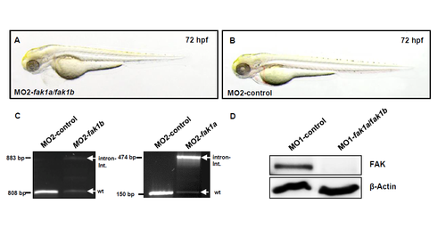
Co-injection of MO2-fak1a and MO2-fak1b results in defective splicing and heart failure in vivo. (A, B) Lateral view of (A) control MO (MO2-control) and (B) MO2-fak1a/fak1b-injected embryos at 72 hpf. The heart failure phenotype of fak1a/fak1b splice morphants was identical to that of embryos injected with the translation blocking FAK MOs (MO1-fak1a/fak1b). (C) RT-PCR of control-, MO2-fak1a- and MO2-fak1b-injected embryos. Injection of MO2-fak1a and MO2-fak1b caused intron integration (808 bp MO2-fak1a; 883 bp MO2-fak1b) leading to premature termination of FAK1a and FAK1b protein translation, respectively. Wild-type fak1a and fak1b RNA was severely reduced in the respective morphants (150 bp MO2-fak1a; 474 bp MO2-fak1b). (D) Western Blot analysis of control and fak1a/fak1b morphant embryos with an antibody against FAK. For each sample 50 embryos were pooled and 20 ?g of protein lysate were loaded per lane. |
|
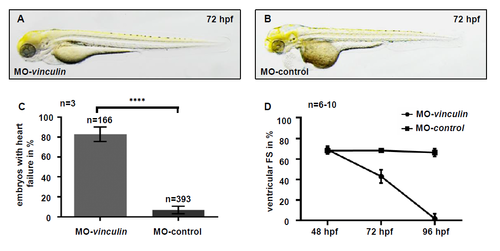
Kockdown of Vinculin results in cardiac contractile dysfunction. (A, B) Lateral view of MO-vinculin (A) and vinculin 5bp-mismatch-MO (MO-control) (B) injected embryos at 72 hpf. (C) Bar graphs compare average of affected embryos after MO injection (MO-vinculin 82.85% ± 6.95%; MO-control 7.3% ± 3.76%; *P<0.0001). (D) Fractional shortening (FS) measurements of Vinculin morphant ventricles compared to control injected embryos at 48, 72 and 96 hpf. FS of Vinculin morphant ventricles was not affected compared to corresponding 5bp-mismatch-MO injected embryos (MO-vinculin 68.01% ± 3.52% vs. MO-control: 66.08% ± 3.12%) at 48 hpf. At 72 hpf, FS in Vinculin morphants was reduced to 42.97% ± 6.53% compared to control morphants (68.26% ± 1.5%), whereas ventricular chambers of Vinculin morphants became almost silent compared to controls at 96 hpf (MO-vinculin: 1.75% ± 4.95%; MO-control: 66.33% ± 3.87%). PHENOTYPE:
|


