- Title
-
Expression change in Angiopoietin-1 underlies change in relative brain size in fish
- Authors
- Chen, Y.C., Harrison, P.W., Kotrschal, A., Kolm, N., Mank, J.E., Panula, P.
- Source
- Full text @ Proc. Biol. Sci.
|
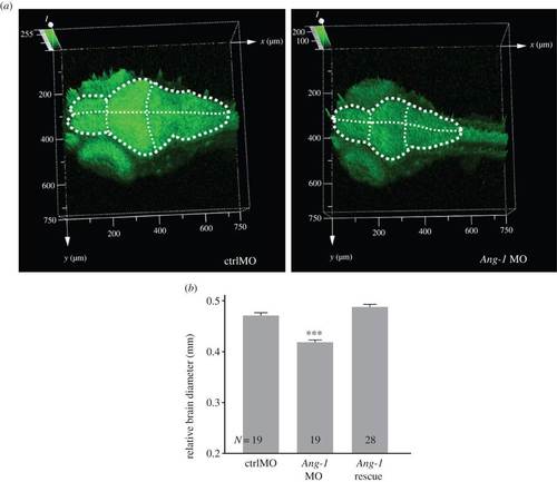
Brain morphology of zebrafish Ang-1 morphant. (a) Knockdown of Ang-1 expression caused the small-sized brain and the reduction of GFP intensity in the Ang-1 morphant (Ang-1 MO) compared with the control group (ctrlMO) in the Tg (alpha-tubulin-GFP) background line at 4 dpf (n = 6). Dashed lines illustrate the brain area. (b) Ang-1 MO shows the significantly smaller relative brain diameter (controlled for body size) than the control and rescue group (ANCOVA: group: F2,62 = 74.55, p < 0.001, body length: F1,62 = 14.47, p < 0.001; post hoc pairwise group comparisons: ctrlMO versus Ang-1 MO: p < 0.001, ctrlMO versus Ang-1 rescue: p = 0.069, Ang-1 MO versus Ang-1 rescue: p < 0.001; ***p < 0.001). PHENOTYPE:
|
|
Notch-1a mRNA expression. (a) Notch-1a expression is dramatically increased in 2-dpf Ang-1 MO morphant brains and 6-dpf brains in the pallium, thalamus, medial and later domains of tectum opticum and intermediate and caudal hypothalamus (n = 6?8 each group). The Ang-1 mRNA normalizes the overexpression of Notch-1a in Ang-1 MO morphants. (b) qPCR analysis of Notch-1a transcript levels (*p < 0.05, n = 9, one-way ANOVA with Dunnett′s test). DT, dorsal thalamus; Hc, caudal hypothalamus; Hi, intermediate hypothalamus; OB, olfactory bulb; P, pallium; RL, rhombic lip; TeO, tectum opticum; VT, ventral thalamus. Scale bar, 100 Ám. |
|
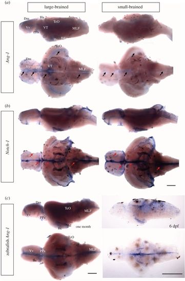
Ang-1 and Notch-1 expression in the guppy fry brains. (a) Expression of guppy Ang-1. (b) Expression of guppy Notch-1. Both Ang-1 and Notch-1 mRNA are present in the medial zone of dorsal telencephalic area (Dm), the ventral nucleus of ventral telencephalic area (Vv), parvocellular preoptic nucleus (PPa), habenula (Ha), ventral thalamus (VT), caudal zone of periventricular hypothalamus (Hc) and dorsal zone of periventricular hypothalamus (Hd), tectum opticum (TeO) and medial longitudinal fascicle (MLF). (c) Ang-1 expression pattern in one-month-old and 6-dpf zebrafish brain. Zebrafish Ang-1 expression is present in Dm, Vv, PPa, Hc, Hd, TeO and MLF. Black and red arrows, respectively, indicate Ang-1 and Notch-1 mRNA difference between the large-brain and the small-brain guppy populations. Scale bars, 200 Ám. EXPRESSION / LABELING:
|


