- Title
-
In vivo imaging of hedgehog pathway activation with a nuclear fluorescent reporter
- Authors
- Mich, J.K., Payumo, A.Y., Rack, P.G., Chen, J.K.
- Source
- Full text @ PLoS One
|
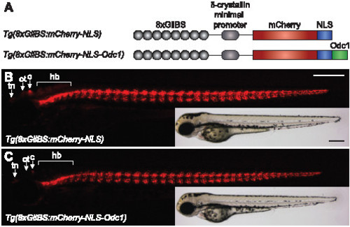
Generation of zebrafish with nuclear Hh pathway reporters. (A) Design of the Gli-dependent nuclear mCherry reporter constructs. (B?C) Tg(8xGliBS:mCherry-NLS) and Tg(8xGliBS:mCherry-NLS-Odc1) larvae at 84 hpf. Fluorescence and brightfield images of representative zebrafish are shown, and the telencephalic nuclei (tn), optic tectum (ot), cerebellum (c), and hindbrain (hb) are labeled. Embryo orientations: lateral view and anterior left. Scale bars: 200 μm. |
|
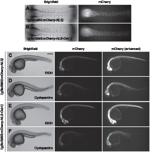
Tg(8xGliBS:mCherry-NLS-Odc1) zebrafish exhibit enhanced reporter turnover. (A?B) Brightfield and fluorescence images of 10-somite (~14 hpf) Tg(8xGliBS:mCherry-NLS) and Tg(8xGliBS:mCherry-NLS-Odc1) embryos. (C?F) The transgenic lines at 30 hpf after the addition of cyclopamine or vehicle control at the 10-somite stage. The embryos were concurrently treated with phenylthiourea (0.003%, w/v) to block pigmentation. Brightness-enhanced fluorescence images are also shown to more clearly depict mCherry-positive cells in cyclopamine-treated embryos. The zebrafish embryos in (A) and (B) are the same animals shown in (D) and (F), respectively. Embryo orientations: A?B, dorsal view and anterior left; C?F, lateral view and anterior left. Scale bars: A?B, 100 μm; C?F, 200 μm. |
|
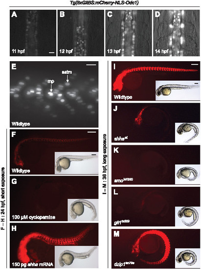
Tg(8xGliBS:mCherry-NLS-Odc1) zebrafish report Hh pathway activity with high sensitivity and fidelity. (A?D) Videomicroscopy frames of a Tg(8xGliBS:mCherry-NLS-Odc1) embryo during early somitogenesis. (E) Somitic region of a Tg(8xGliBS:mCherry-NLS-Odc1) zebrafish at 24 hpf, revealing cellular differences in nuclear mCherry fluorescence. Muscle pioneer cells (mp) and superficial slow-twitch muscle fibers (sstm) are labeled. (F?H) Tg(8xGliBS:mCherry-NLS-Odc1) zebrafish treated with cyclopamine or shha mRNA at the one-cell stage. Fluorescence and brightfield images of representative 24-hpf embryos are shown. (I?M) Tg(8xGliBS:mCherry-NLS-Odc1) zebrafish in wildtype or the indicated mutant backgrounds. Fluorescence and brightfield images of representative 36-hpf embryos are shown. The fluorescence micrographs were acquired with longer exposure times than those in panels F?H to enable the detection of low-level reporter activity. Brightfield micrographs show the distinctive you-type phenotypes observed within the same clutch of embryos. Embryo orientations: A?D, dorsal view and anterior up; E?M, lateral view and anterior left. Scale bars: A?E, 20 μm; F?M, 200 μm. |
|
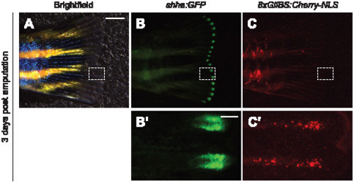
Visualization of Hh signaling during adult fin regeneration. Caudal fin of a Tg(-2.4shha:GFP-ABC;8xGliBS:mCherry-NLS) zebrafish three days after amputation. Brightfield (A) and fluorescence micrographs (B?C) are shown, with the dashed boxes corresponding to the magnified views below. Fin orientations: lateral views, anterior left. Scale bars: A?C, 2 mm; B2?C2, 200 μm. |


