- Title
-
Fam40b is required for lineage commitment of murine embryonic stem cells
- Authors
- Wagh, V., Doss, M.X., Sabour, D., Niemann, R., Meganathan, K., Jagtap, S., Gaspar, J.A., Ardestani, M.A., Papadopoulos, S., Gajewski, M., Winkler, J., Hescheler, J., Sachinidis, A.
- Source
- Full text @ Cell Death Dis.
|
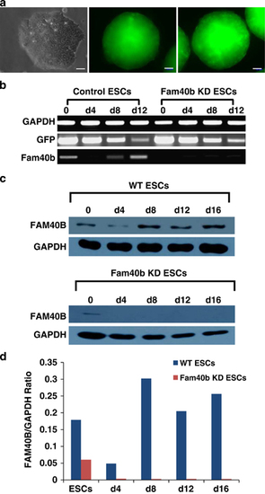
Generation and characterization of ESCs in which Fam40b was constitutively knocked down (KD) by transfection with pGFP-V-RS, expressing shRNA directed against the Fam40b RNA, the GFP and the puromycin resistance cassette. (a) Fluorescence microscopy of control 12-day EBs derived from ESCs transfected with the pGFP-V-RS vector without shRNA (control) and EBs derived from ESCs transfected with the pGFP-V-RS shRNA expressing vector containing 29-mers shRNA (Fam40b KD EBs) (scale bar: 50μm). (b) Semiquantitative RT-PCR analysis of the expression of GFP and Fam40b in undifferentiated control and KD Fam40bESCs as well as in 4-, 8- and 12-day control and KD EBs generated by the hanging drop protocol. (c) Expression of the FAM40B protein during differentiation of WT ESCs (upper panel) and during differentiation of Fam40B KD ESCs. Detection has been performed with FAM40b primary antibody (sc-162799, Santa Cruz Biotechnology; 1:500 dilution) and with the secondary donkey anti-Goat IgG antibody (1:10000 dilution). GAPDH has been detected using the anti-GAPDH antibody (1:25000 dilution). (d) Densitometric analysis of the FAM40B and the corresponding GAPDH bands densities has been performed by using the ImageJ 1.47v software (National Institutes of Health, Bethesda, MD, USA) and the ratio of the FAM40B/GAPDH band densities has been blotted for the different time points of differentiation |
|
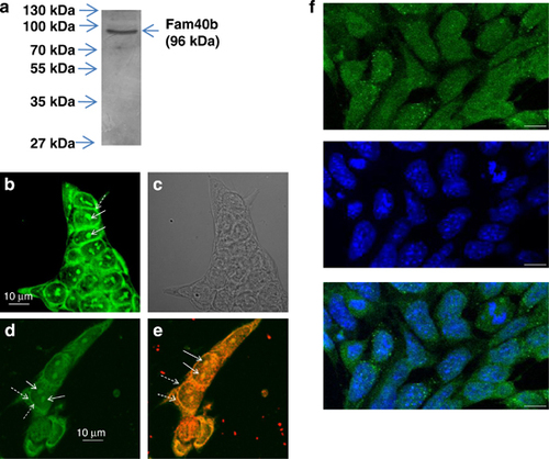
Molecular weight and cellular localization of FAM40B protein. (a) Protein lysates were prepared from undifferentiated ESCs. After separation of 40μg protein by SDS polyacrylamide (10%) gel electrophoresis (SDS-PAGE), western blotting of the proteins was done on nitrocellulose membrane. Chemiluminescence detection of FAM40B has been performed using the Fam40b-433–450 polyclonal antibodies and anti-Mouse IgG alkaline phosphatase-conjugated secondary antibodies. (b and d) Localization of FAM40B in ESCs. ESCs were transfected with the HaloTag Flexi Vector containing the Fam40b cDNA using TurboFect. After 48h, Fam40b was detected using the HaloTag Oregon Green ligand in the nucleoli by confocal microscopy. Normal arrows show Fam40b in the nucleoli and dashed arrows the perinuclear Fam40b. (c) The transparent light microscopy of (b). (e) After fixing of the ESCs (in d), Fam40b has also been detected by immunohistochemistry using primary Anti HaloTag pAb (1:500 dilution) and anti-mouse IgG Alexa Fluor 594 secondary antibodies. (f) Immunostaining of Fam40b in WT ESCs using primary anti-Fam40b antibodies (sc-162799; 1:200) and donkey anti goat IgG-FITC secondary antibody (sc-2024, 1:200) as secondary antibody (upper scan, green pseudocolor). Cells were co-stained with the nuclear marker Hoechst 33342 (scan in the middle, blue). The overlay of nuclear and Fam40b staining (f, bottom) reveals that the presence of Fam40b is not restricted to the nucleus but also extends to perinuclear or even cytoplasmic domains of the ESCs (scale bar: 10μm) |
|
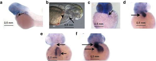
The in situ hybridization of Fam40b in the zebrafish heart as well as the expression of vmhc and of cardiac cmlc2 in control and Fam40b knockdown animals. (a) Digoxigenin-labeled RNA probes were prepared using RNA labeling kit and stained using BM purple. (b) The position of the atrium (arrowhead) and the ventricle (arrow) that was not beating after knockdown of Fam40b is shown. (c) Expression of vmhc in Fam40b knockdown animals was significantly impaired as compared with control animals (d) (arrows show the ventricles). Expression of cmlc2 in Fam40b knockdown animals was almost absent in ventricles and impaired in the atria (e) as compared with control (f) (upper arrows indicate ventricles and lower arrows indicate atria; one representative experiment out of five independent). Whole mount embryos were imaged on a Leica stereomicroscope fitted with a Zeiss Axiocam color camera |


