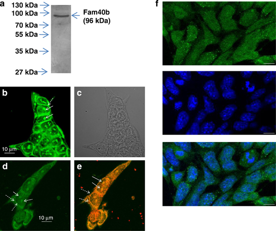
Fig. 6
Molecular weight and cellular localization of FAM40B protein. (a) Protein lysates were prepared from undifferentiated ESCs. After separation of 40μg protein by SDS polyacrylamide (10%) gel electrophoresis (SDS-PAGE), western blotting of the proteins was done on nitrocellulose membrane. Chemiluminescence detection of FAM40B has been performed using the Fam40b-433–450 polyclonal antibodies and anti-Mouse IgG alkaline phosphatase-conjugated secondary antibodies. (b and d) Localization of FAM40B in ESCs. ESCs were transfected with the HaloTag Flexi Vector containing the Fam40b cDNA using TurboFect. After 48h, Fam40b was detected using the HaloTag Oregon Green ligand in the nucleoli by confocal microscopy. Normal arrows show Fam40b in the nucleoli and dashed arrows the perinuclear Fam40b. (c) The transparent light microscopy of (b). (e) After fixing of the ESCs (in d), Fam40b has also been detected by immunohistochemistry using primary Anti HaloTag pAb (1:500 dilution) and anti-mouse IgG Alexa Fluor 594 secondary antibodies. (f) Immunostaining of Fam40b in WT ESCs using primary anti-Fam40b antibodies (sc-162799; 1:200) and donkey anti goat IgG-FITC secondary antibody (sc-2024, 1:200) as secondary antibody (upper scan, green pseudocolor). Cells were co-stained with the nuclear marker Hoechst 33342 (scan in the middle, blue). The overlay of nuclear and Fam40b staining (f, bottom) reveals that the presence of Fam40b is not restricted to the nucleus but also extends to perinuclear or even cytoplasmic domains of the ESCs (scale bar: 10μm)

