- Title
-
Activation of NF-kappaB prevents the transition from juvenile ovary to testis and promotes ovarian development in zebrafish
- Authors
- Pradhan, A., Khalaf, H., Ochsner, S.A., Sreenivasan, R., Koskinen, J., Karlsson, M., Karlsson, J., McKenna, N.J., Orban, L., and Olsson, P.E.
- Source
- Full text @ J. Biol. Chem.
|
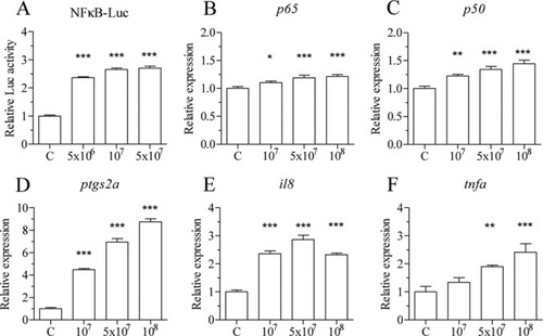
FIGURE 1. Heat-killed bacteria activate NF-?B and inflammatory genes in vitro. nZFL cells were treated with heat-killed E. coli (5 × 10⁶, 1× 10⁷, and 5 × 10⁷ cfu/ml), and the luciferase activity was determined after 12 h of exposure (A). ZFL cells were exposed to heat-killed E. coli (1× 10⁷, 5 × 10⁷, and 1× 10⁸ cfu/ml) for 24 h, and qPCR analysis was performed to determine p65 (B), nfkb1/p50 (C), ptgs2a (D), il8 (E) and tnfa (F). One-way analysis of variance was performed to determine statistical significant difference (*, p < 0.05; **, p < 0.01; ***, p < 0.001). n = 4. Error bars represent the mean ± S.D. |
|
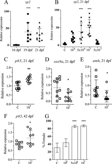
FIGURE 2. Heat-killed E. coli up-regulates p65 and zp2 and leads to a female-biased population. zp2 expression was determined by qRT-PCR from individuals (16, 19, and 21 dpf) using ef1a and 18 S rRNA as reference genes (A). Zebrafish juveniles were exposed to different concentrations of heat-killed bacteria, and zp2 expression was determined by qRT-PCR from individuals exposed from 15?21 dpf (B). Zebrafish juveniles at 20 and 41 dpf were exposed to heat-killed bacteria (107 cfu/ml) for 24 h. Analysis of p65 (C), sox9a (D), and amh (E) expression at 21 dpf juveniles and of p65 (F) expression at 42 dpf gonads was performed by qRT-PCR. Juveniles at 15 dpf were exposed to heat-killed E. coli for 20 days, and their sex was determined at 70 dpf. The mean and S.D. of three independent experiments is shown. Each group contained at least 20 individuals (G). Statistically significant differences between groups were determined using Student's t test for two groups and one-way analysis of variance test for more than two groups (*, p < 0.05; **, p < 0.01; ***, p < 0.001). Error bars represent the mean ± S.D. |
|
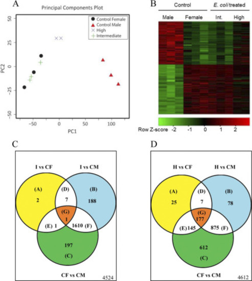
FIGURE 3. The gonadal transcriptomes of individuals treated with heat-killed E. coli are similar to those of control females. Differential gene expression was determined by a homemade gonadal cDNA microarray in the differentiating zebrafish gonads at 35 dpf after treatment with an intermediate (Int; 5 × 10⁶ cfu/ml) and high dose (107 cfu/ml) of heat-killed E. coli. A, shown is a visualization of the main source of experimental variation using principal components analysis of all normalized array expression values. B, shown is a Heatmap visualization of 1812 array features differentially expressed between the control male and control female samples (-fold change ? 2-fold; q value < 0.05). Rows were scaled to have a mean of zero and S.D. of one. C and D, shown is a Venn analysis of genes differentially expressed (-fold change ? 2-fold; q value < 0.05) between the two sexes from the E. coli treatment groups and the control samples, respectively. 1) gene sets are differentially expressed after treatment with intermediate dose of heat-killed bacteria. 2) gene sets are differentially expressed after treatment with high dose of heat-killed bacteria. CF, control female; CM, control male; H, high dose (107 cfu/ml); I, intermediate dose (5 × 10⁶ cfu/ml). The number of genes that were not differentially expressed is indicated in the lower right corner of each panel. |
|
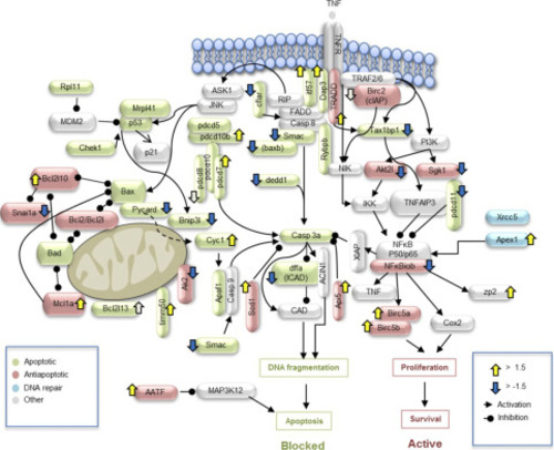
FIGURE 4. Alteration of apoptotic/anti-apoptotic signaling by heat-killed E. coli treatment. Exposure of juvenile zebrafish to an intermediate concentration (5 × 10⁶ cfu/ml) of heat-killed E. coli was performed between 15 and 35 dpf. Gonads were dissected at 35 dpf. After RNA extraction, microarray was used to evaluate apoptotic and anti-apoptotic gene expression in individual gonads. Several critical apoptotic genes were suppressed (cflar, dedd1, pycard, bnip3l, and smac), whereas anti-apoptotic (bcl2l10, aatf, api5, and mcl1a) and NF-?B-target genes (birc5a and birc5b) were up-regulated. |
|
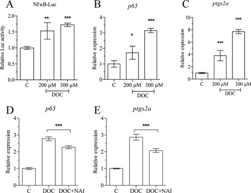
FIGURE 5. DOC activates NF?B and up-regulates p65 and ptgs2a expression in vitro. nZFL were exposed to DOC (200 ?m, 300 ?m) for 12 h, and luciferase activity was analyzed to check NF-?B activation (A). ZFL cells were exposed to DOC (200 ?m, 300 ?m) alone and combination (200 ?m DOC) with NAI (40 nm) for 24 h, and total RNA was extracted followed by qRT-PCR analysis of p65 (B and D) and ptgs2a (C and E) expression. The statistically significant difference between groups was determined using Student's t test (*, p < 0.05; **, p < 0.01; ***, p < 0.001). n = 4. Error bars represent the mean ± S.D. |
|
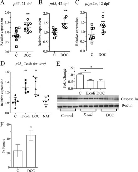
FIGURE 6. DOC treatment up-regulates the expression of inflammatory genes leading to female-biased population. Zebrafish juveniles were exposed to DOC (200 ?m) and analyzed for changes in p65, ptgs2a, and sex ratio. Shown is analysis of p65 levels after 6 days of exposure at 15 dpf (A). 24 h of exposure of 41 dpf juveniles was followed by analysis of p65 (B) and ptgs2a (C) expression in the gonads. Testis explants were exposed to heat-killed bacteria (5 × 10⁷), DOC (200 ?m), and NAI (20 nm) for 24 h, and total RNA was extracted followed by qRT-PCR analysis of p65 (D). Zebrafish juveniles at 70 dpf were exposed to heat-killed bacteria (5 × 10⁷) and DOC (300 ?m) for 2 days, and gonads were isolated for Western blot analysis (E). Larvae at 15 dpf were exposed to DOC (200 ?m) for 20 days, and sex ratio was determined at 70 dpf. The mean and S.D. of two independent experiments are shown. Each group contained at least 30 individuals (F). Statistically significant difference between groups was determined using Student's t test (*, p < 0.05; **, p < 0.01; ***, p < 0.001). Error bars represent the mean ± S.D. |
|
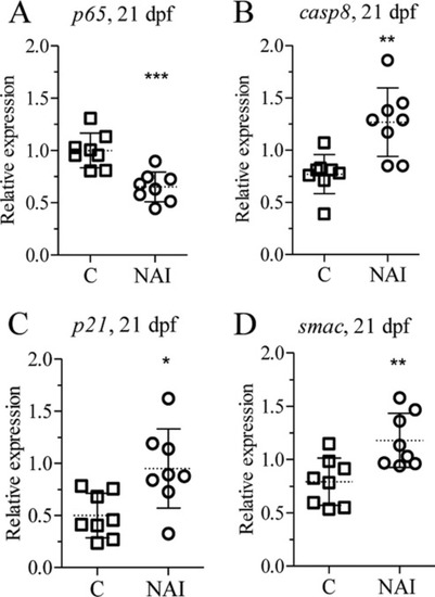
FIGURE 7. NAI exposure alters apoptotic and anti-apoptotic gene expression in vivo. Zebrafish juveniles at 20 dpf were exposed to NAI (20 nm) for 24 h and analyzed for p65 (A), casp8 (B), p21 (C), and smac (D) transcript levels. The statistically significance difference from the control was determined using Student's t test (*, p < 0.05; **, p < 0.01; ***, p < 0.001). n = 8. Error bars represent the mean ± S.D. |
|
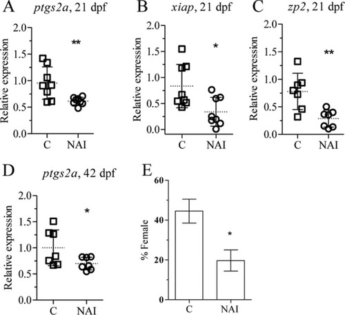
FIGURE 8. NAI treatment down-regulates the expression of inflammatory genes leading to male-biased population. Zebrafish juveniles were exposed to 20 nm NAI for different periods of time followed by qPCR analysis of gene expression and sex ratio. Exposure at 15 dpf for 6 days was followed by analysis of ptgs2a (A), xiap (B), and zp2 (C). Zebrafish juveniles at 41 dpf were exposed for 24 h followed by qPCR analysis of ptgs2a (D). Juveniles at 15 dpf were exposed for 20 days, and the sex ratio was determined at 70 dpf (E). The mean and S.D. of three independent experiments are shown. Each group contained at least 30 individuals. Statistically significance difference from the control was determined using Student's t test (* p < 0.05; **, p < 0.01). Error bars represent the mean ± S.D. |

