FIGURE SUMMARY
Title

Characterization of an lhx1a transgenic reporter in zebrafish

Authors
Swanhart, L.M., Takahashi, N., Jackson, R.L., Gibson, G.A., Watkins, S.C., Dawid, I.B., and Hukriede, N.A.
Source
Full text @ Int. J. Dev. Biol.

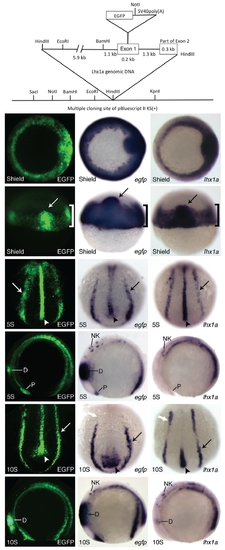
Construct design and early Tg(lhx1a:EGFP)pt303 expression.
(A)
Schematic of lhx1a:EGFP construct. (B-S) Tg(lhx1a:EGFP)pt303 embryos. (B-G) Tg(lhx1a:EGFP)pt303 expression. In situ hybridization for egfp. (N-S) In situ hybridization for lhx1a. (B,H,N) Shield stage embryos, animal view. (C,I,O) Shield stage embryos, dorsal view. Arrow marks the shield and bracket denotes the marginal cells. (D,J,P) 5-somite stage embryos, dorsal-posterior view. Arrow points to intermediate mesoderm and arrowhead marks the notochord. (E,K,Q) 5-somite stage embryos, lateral view. Anterior is to the left. (F,L,R) 10-somite stage embryos, dorsal-posterior view. Arrow points to the intermediate mesoderm and arrowhead marks the notochord. White arrow denotes anterior expression domain of lhx1a in the intermediate mesoderm. (G,M,S) 10- somite stage embryos, lateral view. Anterior is to the left. Abbreviations: D, diencephalon; NK, neural keel; P, polster.

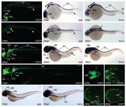
Expression of Tg(lhx1a:EGFP)pt303 during later development.
All animals are oriented such that anterior is to the left. (A-P) Tg(lhx1a:EGFP)pt303 embryos and larvae. ( A , B , G , J , M - P ) Tg(lhx1a:EGFP)pt303 expression. (C,D,H,K) In situ hybridization for egfp. (E,F,I,L) In situ hybridization for lhx1a. (A-F) 24 hpf embryos. Arrow marks position of pronephric proximal tubule and arrowhead delineates distal tubule and duct. (A,C,E) Lateral view, bracket denotes hindbrain. (B,D,F) Dorsal view. (G-I) 48 hpf larvae, arrow in H marks distal tubule and duct in the kidney. (J-L) 72 hpf larvae. (J) Montage of two images to display entire larvae. (M-P) Longitudinal optical sections of transgenic animals. (M) 24hpf embryo. (N-P) 48hpf larva. Abbreviations: aLL, anterior lateral line ganglia; FB, forebrain; HB, hindbrain; MB, midbrain; OV, otic vesicle; pLL, posterior lateral line ganglia; SC, spinal column; TB, tailbud.

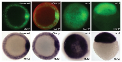
Responsiveness of Tg(lhx1a:EGFP)pt303 to perturbations in Nodal signaling.
(A-H)
Shield stage Tg(lhx1a:EGFP)pt303 embryos. (A-C,E-G) Animal view. (D,H) Lateral view. (A,E) Uninjected control embryos. (B,F) Embryos injected with 125pg mCherry RNA. (C,D,G,H) Embryos injected with 100pg mCherry RNA and 25pg ndr1 RNA. (A-D) Tg(lhx1a:EGFP)pt303 expression. (B) Overlay of mCherry expression (red) displays an embryo that was successfully injected. (E-H) In situ hybridization for lhx1a.

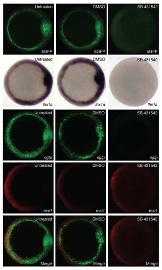
Modulation of Tg(lhx1a:EGFP)pt303 expression by SB-431542.
(A-O)
Shield stage Tg(lhx1a:EGFP)pt303 embryos, animal view. (A,D,G,J,M) Untreated control embryos. (B,E,H,K,N) Embryos treated with 0.8% DMSO. (C,F,I,L,O) Embryos treated with 800 μM SB-431542. (A-C) Tg(lhx1a:EGFP)pt303 expression. (D-F) In situ hybridization for lhx1a. (G-O) Fluorescent in situ hybridization. (G-I) egfp. (J-L) eve1. (M-O) Merged images.

Acknowledgments
This image is the copyrighted work of the attributed author or publisher, and ZFIN has permission only to display this image to its users. Additional permissions should be obtained from the applicable author or publisher of the image. Full text @ Int. J. Dev. Biol.