- Title
-
Differential expression patterns and developmental roles of duplicated scinderin-like genes in zebrafish
- Authors
- Jia, S., Nakaya, N., and Piatigorsky, J.
- Source
- Full text @ Dev. Dyn.
|
A-K: Expression pattern of scinla in the developing zebrafish: 1 day postfertilization (dpf; A,B), 2 dpf(C-E), 3 dpf(F-H), and 6 dpf(I-K). E,H,K: A 10 μm cryosection at the eye level. LE, lens; CO, cornea. |
|
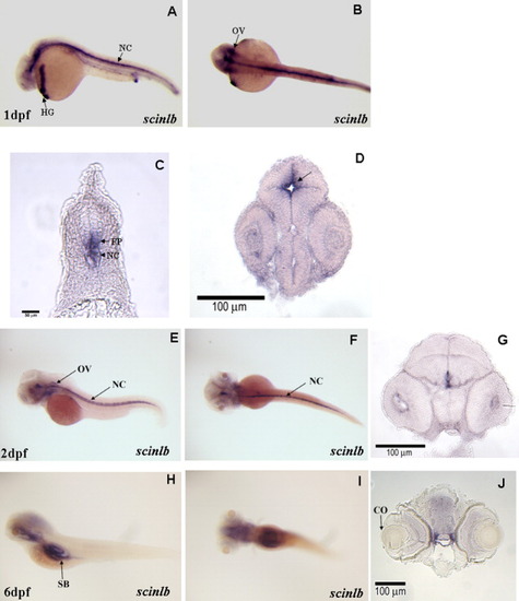
A-J: Expression pattern of scinlb in the developing zebrafish: 1 day postfertilization (dpf; A-D; arrow in D indicates the ventricle of the brain), 2 dpf (E-G), and 6 dpf (H-J). (C,D,G,J) 10 μm cryosection; NC, notochord; HG, hatching gland; OV, otic vesicle; SB, swim bladder; FP, floor plate. |
|
A-C: Enhanced green fluorescent protein (EGFP) expression in the transgenic zebrafish carrying the 12 kb scinlb:EGFP transgene 1 day postfertilization (dpf; A,B) and 3 dpf (C). D: High magnification of the area of square in C. E: Ten micrometer cryosection at the level of a line in C. Blue, nuclear staining with DAPI (4′,6-diamidine-2-phenylidole-dihydrochloride); nc, notochord; hg, hatching gland; e, eye; y, yolk; sc, spinal cord; d, dorsal; v, ventral; r, rostral; c, caudal; *GFP-positive cells at the floor plate. Scale bars = 0.1 mm. EXPRESSION / LABELING:
|
|
Knockdown of scinlb expression by injection of a specific morpholino oligonucleotide (MO). A,B: The scinlb-ATG MO effectiveness assay. C,D: Abnormal development of brain in scinlb-ATG MO microinjected embryos at 29 hours postfertilization (hpf). E,F: Increased cell death in brain and neural tube of scinlb morphant detected by Acridine Orange staining. Arrows represent similar regions. G,H: Decreased expression of shhb in the scinlb morphant. Three separate experiments were conducted, and ∼100 embryos were analyzed each time. EXPRESSION / LABELING:
PHENOTYPE:
|

Unillustrated author statements EXPRESSION / LABELING:
PHENOTYPE:
|


