- Title
-
Regulation of muscle differentiation and survival by Acheron
- Authors
- Wang, Z., Glenn, H., Brown, C., Valavanis, C., Liu, J.X., Seth, A., Thomas, J.E., Karlstrom, R.O., and Schwartz, L.M.
- Source
- Full text @ Mech. Dev.
|
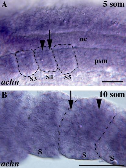
Expression of achn in developing zebrafish somites. (A) Dorsal view of the trunk region at the 5 somite stage (12 h post fertilization (hpf)), anterior left. achn mRNA is expressed at low levels in pre-somitic mesoderm (psm), with more expression medially near the notochord (nc) than laterally. In the first somites that form, achn is expressed in mesenchymal cells (arrowhead), with little expression in epithelialized border cells (arrow). (B) Lateral view of the trunk at the 10 somite stage (15 hpf), anterior left. achn expression is maintained in a mosaic fashion in the mesenchyme (arrowhead), with little expression at somite borders (arrow). Black lines outline somites. Scale bars: 50 μm. EXPRESSION / LABELING:
|
|
Acheron function in zebrafish myogenesis. (A–C) Lateral views of 24 hours postfertilization (hpf) zebrafish embryos labeled with the F59 antibody to reveal slow twitch muscle fibers, white lines show extent of one somite (S). achn mRNA injected embryos (B) had well formed slow fibers (red) and slightly larger somites than seen in uninjected control embryos (A). achn MO injected embryos (C) had reduced somites with less organized slow fibers. (D,F and H) Trunk cross-sections of 48 hpf embryos. achn mRNA injected embryos (F) had larger somites than uninjected embryos (D) with well formed fast fibers throughout the trunk. In contrast, achn MO injected embryos (H) had highly reduced somites with poorly formed fibers. (E,G and I) Confocal images of To-Pro3 stained 48 hpf embryo cross-sections. Left somite is outlined. achn mRNA injection (G) led to a decrease in nuclei. In contrast, ach MO injected embryos (I) had more nuclei despite having smaller somites. (J) Mean diameter (μm) of slow twitch fibers in uninjected, achn mRNA injected, and achn MO-injected embryos. Error bars indicate SEM. (K) Mean number of To-Pro3 labeled nuclei in somite regions of 12 μm sections. Error bars indicate SEM. In A-C scale bar = 20 μm, D–I scale bar = 50 μm. DIC, differential interference contrast microscopy; S, somite. |
Reprinted from Mechanisms of Development, 126(8-9), Wang, Z., Glenn, H., Brown, C., Valavanis, C., Liu, J.X., Seth, A., Thomas, J.E., Karlstrom, R.O., and Schwartz, L.M., Regulation of muscle differentiation and survival by Acheron, 700-709, Copyright (2009) with permission from Elsevier. Full text @ Mech. Dev.


