- Title
-
Cell cycle progression is required for zebrafish somite morphogenesis but not segmentation clock function
- Authors
- Zhang, L., Kendrick, C., Jülich, D., and Holley, S.A.
- Source
- Full text @ Development
|
tiy121 mutants have a cell cycle defect. (A-D) Phosphorylated Histone H3 staining (PHH3, red) marks mitotic cells in wild-type embryos at the shield (A) and 10-somite stage (C). No mitotic nuclei are seen in tiy121 embryos at the shield (B) or 10-somite stage (D). Nuclei are stained with DAPI (blue). (E,F) Wild-type (E) and tiy121-/- (F) embryos at 30 hpf. (G,H) BrdU labeling (red) in the trunks of wild type (G) and tiy121-/- (H) at the 14-somite stage. Asterisks label the notochord. BrdU was injected into the yolk at the 8-somite stage. In C-F, anterior is left; in G and H, anterior is up. PHENOTYPE:
|
|
tiy121 is an emi1 mutant. (A) The chromosomal location of tiy121/emi1. (B) tiy121 is a premature stop codon in emi1. NLS, nuclear localization signal. (C) Graph of the levels of mitosis in tiy121, emi1 morphants, and tiy121 embryos rescued by injection of emi1 mRNA. The level of mitosis, graphed along an arbitrary scale, was wild-type (wt) or reduced to some degree, or mitosis was absent. Morphants and mRNA-injected embryos develop normally prior to the shield stage, thus we infer that the initial level of mitosis is normal without examining PHH3 (dashed lines). hpf, hours post fertilization. (D) tiy121 and hi2648, an insertional allele of emi1, do not complement. (E-G) Expression of emi1 mRNA (E) at the one-cell stage, (F) at the sphere stage, and (G) in the most recently formed somites (arrowheads) and anterior PSM of a 12-somite stage embryo. Anterior is left in G. (H) YFP-Emi1 immunofluorescence. (I) DAPI stained nuclei. (J) Overlay of H and I. Arrows indicate a cell completing mitosis. Scale bar in H: 20 μm for H-J. EXPRESSION / LABELING:
|
|
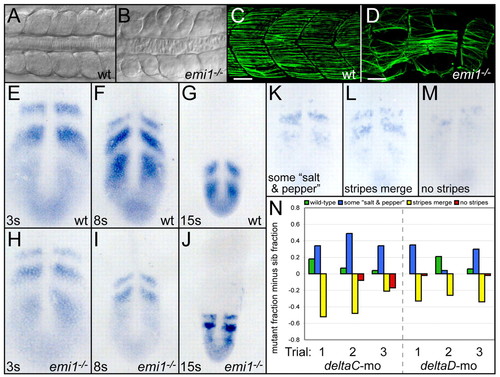
Cell cycle progression is necessary for somitogenesis but not segmentation clock function. (A,B) Dorsal views of anterior trunk somites in wild-type (A) and emi1-/- (B) embryos at the 15-somite stage. (C,D) Posterior trunk myotomes of wild-type (C) and emi1-/- (D) embryos at 36 hpf, lateral views. Slow muscle fibers are labeled with S58 antibodies (green). Scale bars: 30 μm. (E-J) her1 expression at the 3-, 8- and 15-somite stages in (E-G) wild-type and (H-J) emi1-/- embryos. (K-N) her1 stripe integrity was examined in emi1-/- and sibling embryos injected with morpholinos against either deltaC or deltaD. her1 expression was rated according to four categories representing increasing levels of disorganization: wild type; (K) stripes with some `salt and pepper' expression; (L) stripes begin to merge; and (M) no stripes. (N) Distributions of gene expression patterns are displayed for three independent trials (x-axis). Within each gene expression category, the fraction of sibling embryos is subtracted from the fraction of mutant embryos. For example, the wild-type category in the first deltaC morpholino trial included 0.20 fraction of the mutant embryos (20%) and 0.02 fraction of the sibling embryos (2%), giving a graphed value of 0.18. In deltaC morpholino trials, the number of mutants and siblings assayed (mutant/siblings) were: 39/74, 27/79 and 16/57. For deltaD morpholino trials, the corresponding numbers were: 60/50, 28/74 and 49/90. Given the subjective nature of the expression classification, a second assayer performed an independent blind classification of the same embryos (see Fig. S1 in the supplementary material). Although the profiles of the distributions differ, the distinction between emi1 and sibling embryos was consistent. In A-D, anterior is left. In E-M, anterior is up. EXPRESSION / LABELING:
|
|
Cell cycle inhibition leads to somite hyperepithelialization. (A-C) Integrin α5-GFP (red) labels the cell cortex and clusters along somite borders in (A) wild-type (wt), (B) emi1-/- and (C) aphidicolin-hydroxyurea-treated embryos. Embryos were at the 12-somite stage. Somites 2-4, 3-5 and 3-5 are shown, respectively. (D,E) 12-somite-stage emi1-/- embryo labeled with membrane-localized cherry (red) to visualize the cell cortex. The same somites at the beginning (SI and SII) and end (SIV and SV, respectively) of a timelapse are shown. The somite borders are indicated by dashed lines. Somites initially have internal mesenchymal cells, but after three somite cycles have passed, the internal mesenchymal cells (asterisk in D) have moved to the surface of the somite (asterisk in E). (F,G) Fibronectin (Fn) matrix (red) forms along the borders in (F) wild type and (G) emi1-/-. Embryos were at the 12-somite stage. Somites 6-7 and 3-5 are pictured. (H,I) Ephrin B2 expression (green) shows a graded, segmental distribution in (H) wild type and (I) emi1-/-. The lateral membranes of the posterior (p) somite border cells show higher levels of Ephrin B2 than do the lateral membranes of anterior (a) border cells. Embryos were at the 12-somite stage. Shown are somites 5-6 and 4-5. (J,K) Segmental expression of myod (blue) in (J) wild type, somites 3-8, and (K) emi1-/-, somites 4-9, in 10-somite-stage embryos. (L,M) Expression of deltaC (green) in (L) wild type, somites 4-9, is aberrant, but segmental in (M) emi1-/-, somites 4-9, in 10-somite-stage embryos. Nuclei are labeled with DAPI (blue) in A-C,F-I. In D and E, nuclei are labeled with nuclear-GFP (green). β-catenin labels the cell cortex in J,K (yellow) and L,M (red). In all panels, anterior is up. EXPRESSION / LABELING:
|
|
Somite morphogenesis, but not somite polarity, is aberrant in emi1 mutant embryos. (A,B) Lateral views of (A) wild-type and (B) emi1-/- embryos at the 16- to 18-somite stage. (C,D) Lateral view of (C) deltaC morpholino-injected emi1-/- embryos and (D) deltaD morpholino-injected emi1-/- embryos at the 14- to 15-somite stage. (E) An aphidicolin/hydroxyurea-treated embryo at 30 hpf. (F) her1 stripe integrity was examined in emi1-/- and sibling embryos injected with morpholinos against either deltaC or deltaD. her1 expression was rated according to four categories representing increasing levels of disorganization: wild type, stripes with some salt and pepper expression, stripes begin to merge, and no stripes. Distributions of gene expression patterns are displayed for three independent trials (x-axis). Within each gene expression category, the fraction of sibling embryos is subtracted from the fraction of mutant embryos. For example, the wild-type category in the first deltaC morpholino trial included 0.28 fraction of the mutant embryos (28%) and 0.03 fraction of the sibling embryos (3%), giving a graphed value of 0.25. In deltaC morpholino trials, the number of mutants and siblings assayed (mutant/sib) are: 39/74, 27/79 and 16/57. For deltaD morpholino trials, the corresponding numbers are: 60/50, 28/74 and 49/90. These embryos were assayed in Fig. 3N, but here they were examined by a second investigator in a blind trial. (G,H) mesogenin expression in the tailbud of (G) wild-type and (H) emi1-/- embryos at the 15-somite stage. The expression domain is reduced in the mutant. (I,J) mespb expression in the anterior PSM of (I) wild-type and (J) emi1-/- embryos at the 12-somite stage. (K,L) ripply1 expression in (K) wild-type and (L) emi1-/- embryos at the 12-somite stage. (M,N) tbx18 expression in (M) wild-type and (N) emi1-/- embryos at the 12-somite stage. Segmental expression of mespb, ripply1 and tbx18 is established in the mutant. In G-N, anterior is up. (O) Ephrin B2 localization in two wild-type and two emi1-/- embryos. Shown is the 2-9 somite region of 12-somite-stage embryos. Anterior is up. In both mutant and wild type, Ephrin B2 staining is stronger in the cells anterior to each somite border than in the row of cells posterior to each border. EXPRESSION / LABELING:
PHENOTYPE:
|


