- Title
-
Completion of meiosis in male zebrafish (Danio rerio) despite lack of DNA mismatch repair gene mlh1
- Authors
- Leal, M.C., Feitsma, H., Cuppen, E., França, L.R., and Schulz, R.W..
- Source
- Full text @ Cell Tissue Res.
|
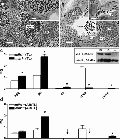
Testis histology of mlh1 -/- zebrafish. a, b Cross section of seminiferous tubule in mlh1+/+ and mlh1-/- zebrafish, respectively, showing spermatogenic cysts with various types of germ cells: different types of spermatogonia (spg), primary spermatocytes (ps), secondary spermatocytes (ss), spermatids (st), spermatozoa (sperm), and apoptotic cells (A). Note the abnormal spermatogenesis in the mutant with many secondary spermatocytes having been released into the lumen, and very few sperm being produced (b, inset, arrow), whereas numerous spermatozoa are visible in the tubular lumen and efferent ducts of wild-type males (a, inset). Bars 25 μm (a, b), 10 μm (insets) c Morphometric analysis of testis sections from fish with TL background. The mutant shows significantly higher amounts of spermatogonia (spg), primary spermatocytes (ps), secondary spermatocytes (ss), and apoptotic cells (apop) than the wild-type. On the other hand, spermatids and spermatozoa (st/sp) are significantly reduced. Inset: Western blot of testes proteins stained with antibodies for MLH1 (85 kDa) and tubulin (50 kDa). MLH1 protein is completely absent in mlh1 -/- testes (-/-) but detectable in wild-type (+/+) and heterozygous (+/-) males. d Morphometric analysis of testis sections from wild-type and mlh1-/- mutant zebrafish on an AB/TL background (n = 6 for both genotypes). The mutant shows an increased mass of spermatogonia (not significant), primary spermatocytes, and apoptotic cells. Secondary spermatocytes, spermatids, and spermatozoa were completely absent (arrows). Significant differences between wild-type and mutant genotypes are indicated (*P < 0.05) PHENOTYPE:
|
|
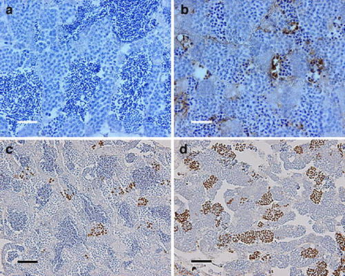
Testis immunohistochemistry of mlh -/- zebrafish. a, b TUNEL staining of mlh1 +/+ and mlh1 -/- fish, respectively. Large numbers of apoptotic cells can be seen in mutant testis (brown). Wild-type testis exhibits a low incidence of apoptosis. c, d Histone H3 staining of cells in metaphase from mlh1 +/+ and mlh1 -/- fish, respectively. Note the higher incidence of stained cells in mutant testis indicating a higher number of mitotic and meiotic cells in metaphase. Bars 25 &mum (a, b), 50 μm (c, d) PHENOTYPE:
|
|
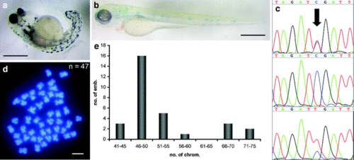
Progeny of mlh1 -/- males. a Severely malformed 3-day-old embryo from a cross of a mlh1 -/- male with a wild-type female. b Unpigmented 3-day-old embryo from a cross of a mlh1 -/- male with an albino female, showing that the wild-type paternal allele of the albino gene has been lost in this embryo. The weak blue staining is caused by the methylene-blue-containing medium. c Sequence traces of the polymorphism in mlh1 (arrow). The paternal allele (mutant) is a T, and the maternal allele (wild-type) is a C. The sequence peak areas show quantitatively that some progeny of mutant males have one chromosomal copy of each parent (top), whereas others have only the maternal allele (middle) or two copies of the paternal allele (bottom that some progeny of mutant males have one chromosomal copy of each parent (top), whereas others have only the ma). d Embryos have chromosome numbers different from the normal number of 50, such as this example of 47 chromosomes. e Distribution of chromosome numbers in embryos (emb.) from mlh1 -/- males (30 embryos in total). Bars 500 μm (a, b), 1 μm (d) PHENOTYPE:
|

Unillustrated author statements PHENOTYPE:
|


