- Title
-
The zebrafish, brain-specific, aromatase cyp19a2 is neither expressed nor distributed in a sexually dimorphic manner during sexual differentiation
- Authors
- Kallivretaki, E., Eggen, R.I., Neuhauss, S.C., Kah, O., and Segner, H.
- Source
- Full text @ Dev. Dyn.
|
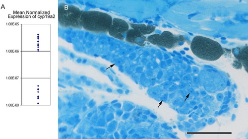
cyp19a2 expression in the head of 20 days postfertilization (dpf) fish and histological evaluation of gonadal development. A: Mean normalized expression (MNE) of cyp19a2 expression in 20 dpf zebrafish. There are clearly two populations, namely the high and low expressing fish. B: Histological evaluation of the gonadal status of these fish. The germ cells are represented mainly by gonial cells (arrows) possessing a rather homogeneous, basophilic cytoplasm with usually one, centrally located nucleolus; the cytoplasm is restricted to a relatively small rim around the nucleus. These germ cells will probably differentiate into oocytes, because the gonads of juvenile zebrafish initially develop as nonfunctional protogynic ovaries (Maack and Segner, [2003]). However, because oocytes are not yet present, these gonads were classified as undifferentiated. Scale bar = 50 μm. EXPRESSION / LABELING:
|
|
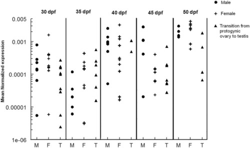
Ontogeny of cyp19a2 mRNA levels in the brains of juvenile zebrafish. Fish were collected throughout the process of gonadal differentiation (30, 35, 40, 45, 50 dpf), and the heads were used for RNA extraction and quantification of cyp19a2 transcript abundance, while the bodies were kept for histological evaluation of gonadal status. Males are represented as black filled circles, females as crosses, and animals undergoing transition from stage I oocyte to testis as black filled triangles. At 50 dpf, transcript levels were significantly higher compared with all other stages (Wilcoxon test). The results are expressed as mean normalized expression (MNE; Muller et al., [2002]). Gene levels were normalized against 18S. EXPRESSION / LABELING:
|
|
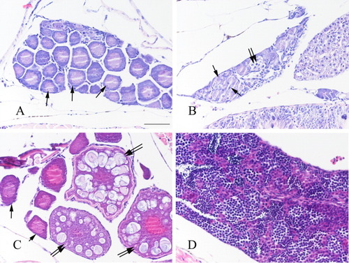
Gonadal histology of sexually differentiating zebrafish. A: Early ovary containing only immature stage I oocytes of the perinucleolar stage (arrows). B: Early ovary undergoing transition to testicular tissue, with degenerating oocytes (arrows) and spermatogonial cysts (double arrow). C: Progressed ovary with perinucleolar (arrows) and cortical alveolar (double arrows) oocytes. D: Testis. Scale bar = 50 μm. |
|
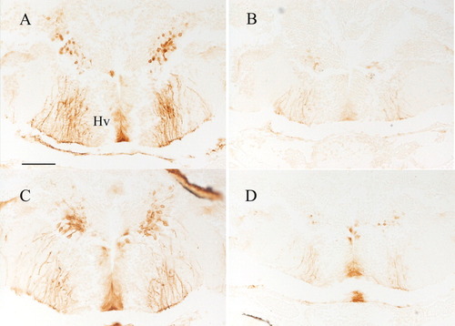
Differences in CYP19A2 staining intensity among female and male 40 days postfertilization (dpf) zebrafish. Both micrographs depict the area surrounding the ventral zone of the periventricular hypothalamus. A,C: High expression of aromatase in a female (A) and male (C) fish. B,D: Low expression in a female (B) and male (D) 40 dpf fish. Scale bar = 50 μm. EXPRESSION / LABELING:
|
|
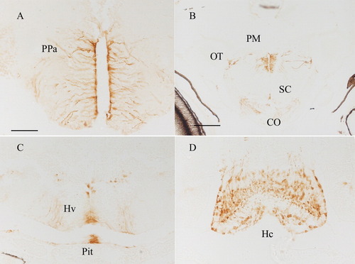
CYP19A2-immunoreactive cells in the telencephalon (A,B) and diencephalon (C,D) of 40 days postfertilization (dpf) zebrafish. A: Parvocellular preoptic nucleus (PPa). B: Magnocellular preoptic nucleus (PM)/optic chiasm (CO)/optic tract (OT)/suprachiasmatic nucleus (SC). C: Ventral zone of periventricular hypothalamus (Hv)/pituitary (Pit). D: Caudal zone of periventricular hypothalamus (Hc). Scale bar = 50 μm. EXPRESSION / LABELING:
|
|
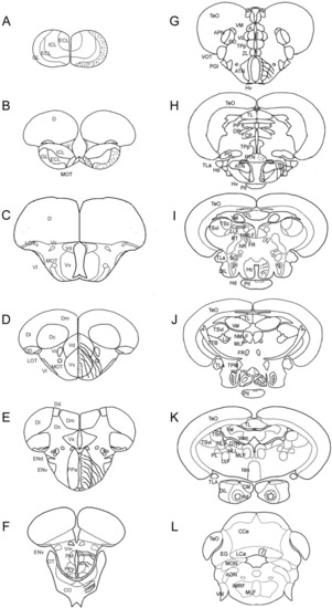
A-K: CYP19A2 immunoreactivity in the telencephalon (A-F), diencephalon (G-K) and cerebellum of 40 days postfertilization (dpf) and 50 dpf zebrafish. GL, glomerular layer of olfactory bulbs; MOT, medial olfactory tract; Vv, ventral nucleus of ventral telencephalic area; Vd, dorsal nucleus of ventral telencephalic area; PPa, parvocellular preoptic nucleus anterior part; OT, optic tract; PM, magnocellular preoptic nucleus; PPp, parvocellular preoptic nucleus posterior part; VM, ventromedial thalamic nucleus; SC, suprachiasmatic nucleus; CO, optic chiasm; ZL, zona limitans; ATN, anterior tuberal nucleus; PTN, posterior tuberal nucleus; Hv, ventral zone of periventricular hypothalamus; Hc, caudal zone of periventricular hypothalamus; Hd, dorsal zone of periventricular hypothalamus; Pit, pituitary; LCa, caudal lobe of cerebellum. EXPRESSION / LABELING:
|
|
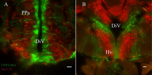
Immunofluorescence staining with anti-CYP19A2 and anti-HuC/D. A: Detail from the anterior part of the parvocellular preoptic nucleus (PPa). CYP19A2 stains the cells lining the diencephalic ventricle (DiV), whereas anti-HuC/D stains the surrounding neurons. B: The anti-CYP19A2 antibody stains cell bodies in the area surrounding the ventral zone of the periventricular hypothalamus (Hv), and their processes that run down to the border of the brain that comes in contact with the pituitary (Pit). The cell bodies that are stained within the ventral zone of the periventricular hypothalamus (Hv) differ morphologically from the rest in the surrounding area as they are more elongated. Scale bar = 10 μm in A, 15 μm in B. EXPRESSION / LABELING:
|
|
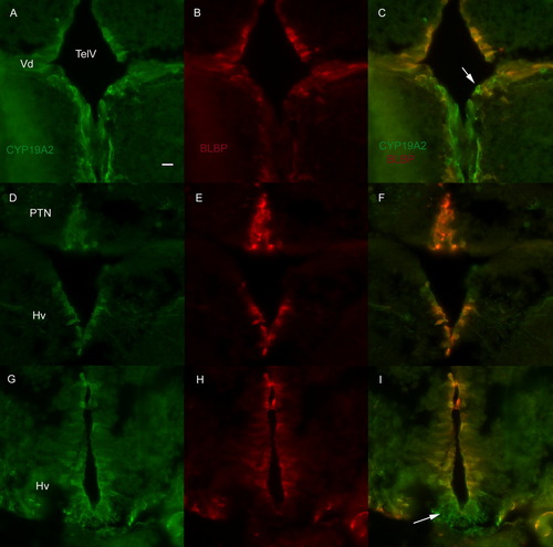
Double immunofluorescence for CYP19A2 and BLBP. A-C: Most of the cells that surround the telencephalic ventricle (TelV) at the level of the dorsal nucleus (Vd) of the ventral telencephalic area that are positive for CYP19A2 are also positive for BLBP, with some exceptions (arrows). D-F: The majority of the cells in the posterior tuberal nucleus (PTN) and at least some levels of the ventral zone of the periventricular hypothalamus (Hv) are double positive. G-I: The part of the Hv that comes in contact with the pituitary is not double labeled (arrow in I). Scale bar = 10 μm. EXPRESSION / LABELING:
|

