- Title
-
Completing the set of h/E(spl) cyclic genes in zebrafish: her12 and her15 reveal novel modes of expression and contribute to the segmentation clock
- Authors
- Shankaran, S.S., Sieger, D., Schroter, C., Czepe, C., Pauly, M.C., Laplante, M.A., Becker, T.S., Oates, A.C., and Gajewski, M.
- Source
- Full text @ Dev. Biol.
|
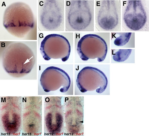
Expression pattern analysis of her15 in the caudal PSM. Expression of her15 was analysed from 70% epiboly up to 16-somite stage focusing on the caudal PSM. Expression first appears during epiboly at the epibolic margin while the dorsal midline is devoid of transcript (A and B). Note expression on either side of the dorsal midline extents during epiboly towards the anterior pole (white arrow). At bud an expression domain is observed posterior to the notochord, which shows different width in a batch of embryos of nearly the same developmental stage (C?F). Due to narrowing and lengthening of the tail bud only broad and dot-like expression can unambiguously be distinguished in 12- to 14- (G and H, respectively) or 16- to 18-somite stage embryos (I and J, respectively; K and L are magnifications of the tail bud in panels I and J). During these stages it also becomes evident that the staining in the dot-like expression phase extends from ventral to dorsal in the midline of the tail bud (H and J). Wt embryos at the 10- to 12-somite stage were double-stained for her7 transcripts (red) and her15 (M, N) or her12 (O, P) transcripts (blue). ?Broad? staining patterns for her15 and her12 coincided with ubiquitous her7 expression in the posterior PSM (M, O), ?dot-like? her15 and her12 patterns were associated with low her7 staining in the this region (N, P). Arrowhead in panel P points to a region in the caudal PSM with her12 transcription only, indicating slight phase shifts to her7. EXPRESSION / LABELING:
|
|
Expression of the MMLV-retroviral enhancer trap CLGY-521 during somitogenesis. (A) Schematic representation of the CLGY-521 insertion site 535 bp 5′ to the her15b gene on Chr 11. LTR = long terminal repeat, gata2P = zebrafish gata2 promoter, YFP = yellow fluorescent protein. (B, B′) Representative embryos from weak and strong expression classes, respectively, showing yfp mRNA distribution at bud stage; vegetal views, dorsal up. (C, C′) Weak and strong expression classes at 12 somites, dorsal view of tail bud. (D, D′) Same embryos in panels C, C″ viewed laterally, to highlight the ventral core of expression. (E) Embryo from panels C, D viewed axially from posterior, to reveal the ring-like shape of expression domain. (F, F′) Weak and strong expression classes at 17 somites, lateral view of tail bud. |
|
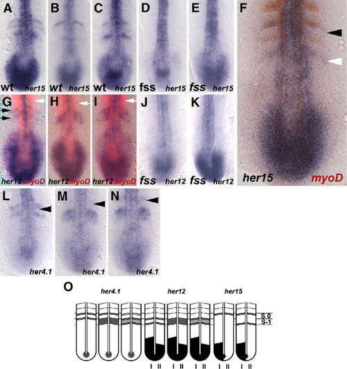
Expression pattern analysis of the mHes5 homologues in the rostral PSM. (A?C, F) wt expression pattern of her15. At 8- to 10-somite stage her15 expression was observed in one to two thin stripes in the rostral half PSM (A?C). These stripes occur in a double segmental distance at the future posterior somite borders of S1 and S-1, or when compared to myoD are co-expressed with the last myoD stripe and the myoD stripe within the last forming or formed somite (F). (D, E) her15 expression in fss. (G?I) wt expression pattern of her12 (blue) double-stained with myoD (red). (J, K) her12 expression in fss. (L?N) wt expression pattern of her4.1. In contrast to her15, the stripes of her12 and her4.1 expression occur in a single segmental distance in the rostral PSM (G?I and L?N, respectively). (F) wt expression of her15 (in blue) compared to myoD (in red). Black arrowhead points to the anterior-most her15 and her4 expression stripe, respectively, at the forming segment border. White arrowhead marks the posterior her15 stripe, which is located in the last myoD stripe. Black arrow indicates the position of the two her12 stripes matching with the penultimate and ultimate myoD stripe, respectively. White arrow marks the last formed segment border. (O) Schematic representation of the expression stripes of the mHes5 homologues in the rostral PSM. Location of the striped expression in the rostral PSM is compared between her4.1, her12 and her15. Note, cyclic expression in the posterior is indicated by roman numbers I and II below the respective drawing. S0 = somite, which will be formed next; S-1 = prospective future somite posterior to S0. EXPRESSION / LABELING:
|
|
Delta?Notch control of her12 and her15 expression. her12 expression was examined in Su(H) morphants (2 experiments, n = 125, 96% affected), after DAPT treatment (2 experiments, n = 41, 98% affected), in the fused somite type mutants aei/deltaD, bea/deltaC, des/notch1a and in her7 morphants (2 experiments, n = 93, 85% affected) and compared to the respective wild type expression. Similarly, her15 expression was analysed in Su(H) morphants (3 experiments, n = 21, 100% affected), after DAPT treatment (2 experiments, n = 37, 98% affected), in the fused somite type mutants aei/deltaD, bea/deltaC, des/notch1a and in her7 morphants (2 experiments, n = 74, 69% affected) and compared to the respective wild type expression. Wild type (wt) expression of her12 and her15 (A and H, respectively). Expression of her12 and her15 in Su(H) morphants (B and I), after DAPT treatment (C and J), in aei (D and K) in bea (E and L) and in des (F and M, respectively) at the 10- to 12-somite stage. (G and N) her12 and her15 expression in her7 morphants, respectively. (O?R) (S?V) her15 expression in aei, bea, des and Su(H) morphants at the 12- to 14-somite stage (in lateral view) and at bud stage, respectively. White arrowhead points to the ventral expression domain of her15 that is only lost in the aei mutant situation. EXPRESSION / LABELING:
|
|
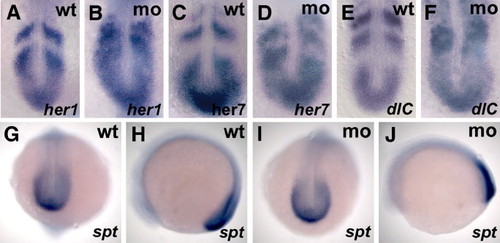
Influence of the her12?ORF-Mo on gene expression in the PSM. (A, C, E) Wild type expression of her1, her7 and deltaC, respectively. (B, D, F) Expression of her1, her7 and deltaC, respectively, after her12?ORF-Mo injection (0.8 mM; 3 experiments for her1, n = 115, 55% affected; 2 experiments for her7, n = 65, 51% affected; 2 experiments for deltaC, n = 72, 42% affected). (G, H) Wild type expression of spt, (I, J) spt expression in her12 ORF-morphants (0.8 mM, 2 experiments, n = 68, 96% of all embryos show wild type expression). (A?F) Flat-mounted embryos (8- to 10-somite stage), anterior to the top. (G?J) Whole mount embryos (10-somite stage); (G, I) dorsal view, posterior downwards; (H, J) lateral view, anterior to the left. EXPRESSION / LABELING:
|
|
Effects of her12 and her15 over-expression on somite morphology and clock genes. (A-C) GFP-RNA-injected embryos (850 ng/Ál GFP-RNA, 2 experiments, n = 82, 96% show wild type morphology). (D-F) her12 RNA-injected embryos (800 ng/Ál her12-RNA, 3 experiments, n = 102, 44% affected). (G, I, K) GFP-RNA-injected embryos stained for her1, her7 and deltaC, respectively (850 ng/Ál GFP-RNA, 2 experiments, n = 96, 95% show wild type expression). (H, J, L) her12-RNA-injected embryos stained for her1, her7 and deltaC, respectively (800 ng/Ál her12-RNA, 2 experiments, n = 104, 48% affected). (M, O) GFP-RNA-injected embryos (150 ng/Ál GFP-RNA, 4 experiments, n = 235, 95% show wild type morphology). (N, P) her15 RNA-injected embryos (115 ng/Ál, 4 experiments, n = 219, 44% show somite border defects). Note that another 34% of the injected embryos displayed gastrulation defects indicating that her15 might act pleiotropically. In addition misexpression of her15 caused severe brain defects resulting in improper brain patterning, enlarged brain vesicles and eye defects (compare M with N), while her12 misexpression causes overall brain enlargement (compare A, B with D, E, respectively). Compared to GFP-mRNA control injections (Q, R and T, U, respectively) cyclic gene expression of deltaC (S) and her1 (V) is disrupted. (A, B, D, E, M, N) whole mount embryos, lateral view, anterior to the upper left; (C, F, O, P) dorsal view, anterior to the top; (G-L and Q-V) flat-mounted embryos, anterior to the top; (A, C, D, F-L and Q-V) 10-somite stage embryos; (B, E and M-P) 14-somite stage embryos. EXPRESSION / LABELING:
|
|
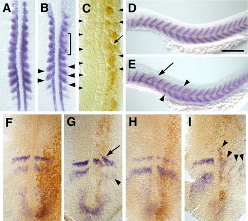
Somitogenic and cyclic expression defects from her7 mRNA over-expression. The expression of myoD in the presomitic mesoderm and trunk somites of embryos at 14 hpf (10 som, A, B) and cycling genes in the presomitic mesoderm at 10 hpf (1 som, F?I) are shown in dorsal view after flat mounting with anterior up. The myotome boundaries of the trunk marked by titin expression are shown in 26 hpf embryos in lateral view, anterior to the left and dorsal up (D, E). Scale bar in panel D is 250 Ám. Arrows and arrowheads indicate localized defects and brackets indicate the extent of larger regions of abnormalities. Expression of myoD after injection of lacZ (A) or her7 mRNA (B). Segmentation defects of the trunk coincide with the region in which the Myc-tagged Her7 protein can be detected (C). Segmentation of the trunk after injection with lacZ mRNA (D) or her7 mRNA (E). Expression of delC (F, G) and her1 (H, I) after the injection of gfp (F, H) or Myc?her7 mRNA (G, I). EXPRESSION / LABELING:
|
Reprinted from Developmental Biology, 304(2), Shankaran, S.S., Sieger, D., Schroter, C., Czepe, C., Pauly, M.C., Laplante, M.A., Becker, T.S., Oates, A.C., and Gajewski, M., Completing the set of h/E(spl) cyclic genes in zebrafish: her12 and her15 reveal novel modes of expression and contribute to the segmentation clock, 615-632, Copyright (2007) with permission from Elsevier. Full text @ Dev. Biol.


