- Title
-
Characterization and expression pattern of two zebrafish atf7 genes
- Authors
- Zhao, C., Qi, J., and Meng, A.
- Source
- Full text @ Dev. Dyn.
|
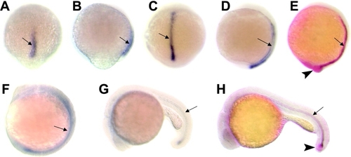
Spatiotemporal expression pattern of zebrafish atf7a. Distribution of endogenous mRNA was detected by whole-mount in situ hybridization. A,B: The 80% epiboly stage. C-E: The bud stage. F: The 10-somite stage. G,H: At 1 day. A,C: Dorsal views with animal pole to the top. B,D,E,F: Lateral views with animal pole or anterior to the top. E,H: Double staining with atf7a (blue) and ntl (red). Arrows indicate the notochord precursors or the notochord. E,H: Arrowheads indicate the germ ring (E) or tail bud (H) where only ntl is expressed. EXPRESSION / LABELING:
|
|
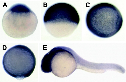
The spatiotemporal expression pattern of zebrafish atf7b. In contrast to atf7a, atf7b is ubiquitously expressed at all stages examined. A: At the one-cell stage, with the animal pole to the top. B: The shield stage, lateral view with dorsal to the right. C: The shield stage, animal-pole view with dorsal to the right. D: The bud stage, lateral view with the dorsal to the right. E: At 24 hours postfertilization, lateral view with anterior to the left. |
|
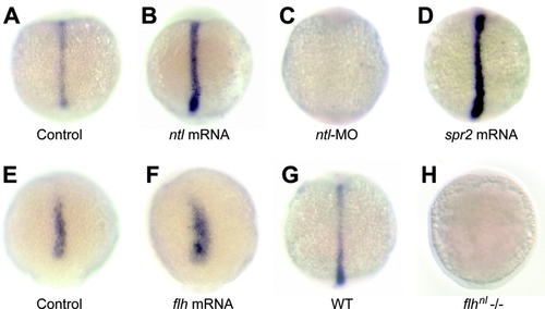
Regulation of atf7a expression by transcription factors Ntl, Spr2, and Flh. A,E: Control embryos injected with green fluorescent protein (GFP) mRNA. B,D,F: Injection with 150 pg of ntl mRNA (B), 100 pg of spr2 mRNA (D), or 100 pg of flh mRNA (F) enhanced atf7a expression. C: Injection with 10 ng of ntl-morpholino inhibited atf7a expression. G,H: The expression of atf7a was not detected in the flhnl mutant embryo (H), whereas it was expressed in the wild-type embryo from the same cross (G). A-D,G,H: The bud stage. E,F: The 90% epiboly stage. All embryos were dorsal views, with the animal pole to the top. EXPRESSION / LABELING:
|


