- Title
-
T-box gene eomesodermin and the homeobox-containing Mix/Bix gene mtx2 regulate epiboly movements in the zebrafish
- Authors
- Bruce, A.E., Howley, C., Dixon Fox, M., and Ho, R.K.
- Source
- Full text @ Dev. Dyn.
|
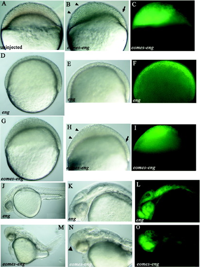
eomes-eng inhibits epiboly. A-O: All views lateral. J-O: anterior toward the left. Construct injected, if any, is indicated in the bottom left corner. A-C: Embryos at 30% epiboly (4.7 hours postfertilization [hpf]), (D-I) embryos at 60% epiboly (6.5 hpf). A: Uninjected control. B: Embryo injected with gfp and eomes-eng RNA. Arrowheads indicate the region of the blastoderm that has failed to thin, and the arrow indicates the normal region of the blastoderm. C: Same embryo as in B showing green fluorescent protein (GFP) fluorescence. The region indicated by the arrowheads in B is where most of the GFP expression is located. D: Control embryo injected with gfp and eng RNA. E: Higher power view of embryo in D. F: Same embryo as in E, showing that GFP fluorescence is distributed throughout the blastoderm. GFP-positive cells are intermingled with unlabeled cells. G: Embryo injected with gfp and eomes-eng RNA. H: Higher power view of embryo in G. Arrowheads indicate region of the blastoderm that has failed to thin, and the arrow indicates the normal region of the blastoderm. I: Same embryo as in H, showing GFP fluorescence. The region indicated by the arrowheads in H is where most of the GFP expression is located. J-O: Embryos at 1 day postfertilization. J: Control embryo injected with gfp and eng RNA. K: Higher magnification of J, showing the head region. L: Same embryo as in K, showing evenly distributed GFP fluorescence. M: Embryo injected with gfp and eomes-eng RNA. N: Higher magnification of M, showing abnormal head region. O: Same embryo as in N showing GFP fluorescence concentrated in the anterior portion of the head. |
|
Eomes is expressed throughout the blastoderm at early blastula stages. Images are single scans from a confocal z-series of embryos shown in lateral view and stained with the anti-Eomes antibody. A: Embryo at 512-cell stage (2.75 hours postfertilization [hpf]). Nuclear staining can be seen throughout the blastoderm, although not all nuclei are in the plane of view. B: Embryo at the high stage (3.3 hpf). Protein expression can be seen in nuclei throughout the blastoderm. EXPRESSION / LABELING:
|
|
eomes regulates mtx2 expression cell-autonomously. All views are lateral, and all embryos are at sphere stage (4 hours postfertilization [hpf]). Injected construct, if any, is indicated in lower left corner. A: In situ hybridization of mtx2 in an uninjected embryo, showing expression in the marginal cells of the blastoderm and the underlying yolk syncytial layer. B: eomes-VP-injected embryo with ectopic mtx2 expression (arrowhead). Inset shows a portion of the blastoderm of a myc-eomes-injected embryo with Eomes protein expression in the nucleus in brown and mtx2 expression in blue. White outline demarcates a group of cells that coexpress Eomes and ectopic mtx2, indicating a cell-autonomous induction of mtx2 by Eomes. C: Reduced mtx2 expression in an embryo injected with eomes-eng. D: ntl-VP-injected embryo with normal mtx2 expression. EXPRESSION / LABELING:
|
|
mtx2 morpholinos inhibit epiboly. All views are lateral with dorsal to the right; all embryos are at shield stage (6 hours postfertilization). Injected construct, if any, is indicated in lower left corner. A: Uninjected embryo. B: Embryo injected with Mtx2-MO1 into one cell at the two-cell stage; note that the blastoderm is thickened compared with control. C: Embryo injected with Mtx2-MO1 into the yolk syncytial layer (YSL); note that the blastoderm is thickened compared with control. |
|
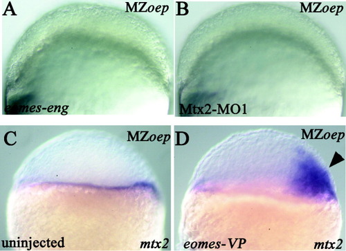
Epiboly defect is Nodal-independent. All views are lateral; the mutant phenotype is indicated in upper right corner; the injected construct is indicated in lower left corner. A,B: At 50% epiboly (5.25 hours postfertilization [hpf]). C,D: At sphere stage (4 hpf). A: Embryo injected with eomes-eng; the blastoderm has failed to thin. B: Embryo injected with Mtx2-MO1; the blastoderm has failed to thin. C: mtx2 expression in an uninjected embryo. D: Ectopic mtx2 expression (arrowhead) in an eomes-VP-injected (into a single cell at the eight-cell stage) embryo. EXPRESSION / LABELING:
|

Unillustrated author statements |


