- Title
-
Characterization of expanded intermediate cell mass in zebrafish chordin morphant embryos
- Authors
- Leung, A.Y., Mendenhall, E.M., Kwan, T.T., Liang, R., Eckfeldt, C., Chen, E., Hammerschmidt, M., Grindley, S., Ekker, S.C., and Verfaillie, C.M.
- Source
- Full text @ Dev. Biol.
|
Morphology of WT (A, 24 hpf; C, 48 hpf) and ChdMO embryos (B, 24 hpf; D, 48 hpf). White arrowhead: Intermediate cell mass. The inserts showed embryos stained with O-dianisidine. Brown color indicates the presence of hemoglobin. |
|
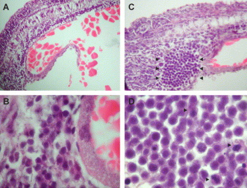
Hematoxylin and eosin sectioning of WT (A, B) and ChdMO embryos (C, D) at low (A, C) and high-power (B, D) magnification at 24 hpf. In WT embryos, the ICM is only 3?5 cell thick posterior to the yolk extension. The ChdMO embryos had expanded ICM (arrowheads in panel C), which contained a monotonous population of cells with regular and round nuclei and high nuclei to cytoplasm (N/C) ratio. Mitotic figures are easily identified (arrowheads in panel D). |
|
H&E sectioning of WT (A, B) and ChdMO embryos (C, D) at low (A, C) and high power (B, D) magnification at 48 hpf. In WT embryos, there was no defined population of cells in the ICM although erythroid cells can be readily identified at the axial circulation (B). In the chordin morphants, the ICM remained expanded but instead of a monotonous population seen at 24 hpf, the cells in ICM at 48 hpf became heterogeneous. Monocyte-looking cells with horseshoe-shaped nuclei are readily encountered (arrowheads). |
|
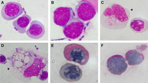
Wright staining of cytospin preparation of cells dissected out from the expanded ICM of the ChdMO embryos at 48 hpf. The ICM comprised heterogeneous population of cells, including erythroid cells (A), blast-looking cells (B), monocyte-looking cells (arrowhead, C and D). Some of the erythroid cells (E) as well as blast-looking cells (F) were PAS (Periodic Acid Schiff) staining positive. |
|
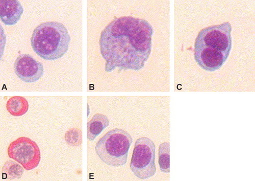
Wright staining of cytospin preparation of cells dissected out from the circulation of the ChdMO and wild-type embryos at 48 hpf. Most of the circulating cells are erythroid in morphology (A), monocytes (B), and binucleated erythroid cells (C) accounted for 1.2 and 1.4 per 200 cells, respectively. Periodic Acid Schiff (PAS) staining showed positivity in 21% of cells (D). (E) Circulating erythroid cells in the WT embryos. 2.0 per 200 cells in the WT embryos were binucleated (not shown). Monocytes were not evident. PAS staining was all negative in WT embryos. |
|
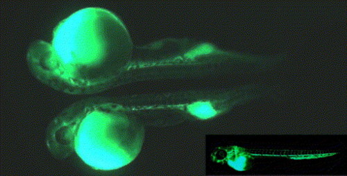
Microangiographic studies in ChdMO embryos at 48 hpf showing that the expanded ICM was in continuity with the rest of the circulation. The insert showed a WT embryo at 48 hpf. |
|
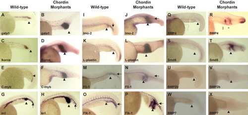
Whole-mount in-situ hybridization of WT and ChdMO embryos at 24 hpf. Expression of both hematopoietic and vascular genes, as well as BMP4 and smad5, were up-regulated in the expanded ICM (arrowhead). In the ventral tail region (arrow), there was no difference in the expression of genes between WT and the morphants. Note that BMP4 was ectopically expressed in the morphants at the tip of the yolk extension (?) as well as the distal border of the expanded ICM (*). EXPRESSION / LABELING:
|
|
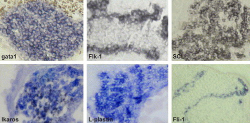
Paraffin sections (7 Ám) of ChdMO embryos (24 hpf) at ICM after hybridization. Gata1, ikaros, l-plastin, and scl were generally expressed among the cells in the ICM. Vascular genes including flk1 and fli1 were expressed at the periphery of the ICM. |
|
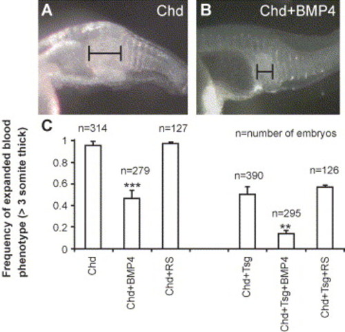
Co-injection of BMP4 MO with chordin MO reduced ICM expansion in chordin morphants. Upper panel: (A?B) ICM expansion in morphant embryos (A) and morphant embryos co-injected with BMP4 MO (B). A width of >3 or d3 somites was arbitrarily defined as the cutoff for ICM expansion. Lower panel: (C) Left: Frequency of ICM expansion in embryos injected with chordin MO (Chd, 0.75 ng), Chd MO (0.75 ng) + BMP4 (12 ng), ChdMO (0.75ng) + random sequence MO (12 ng). Right: Frequency of ICM expansion in embryos injected with ChdMO (0.15 ng) + TsgMO (6 ng), ChdMO (0.15 ng) + TsgMO (6 ng) + BMP4MO (12 ng), ChdMO (0.15 ng) + TsgMO (6 ng) + random sequence MO (12 ng). Each error bar represents 1 SEM. *P < 0.05. RS, random sequence MO (see Materials and methods). |
Reprinted from Developmental Biology, 277(1), Leung, A.Y., Mendenhall, E.M., Kwan, T.T., Liang, R., Eckfeldt, C., Chen, E., Hammerschmidt, M., Grindley, S., Ekker, S.C., and Verfaillie, C.M., Characterization of expanded intermediate cell mass in zebrafish chordin morphant embryos, 235-254, Copyright (2005) with permission from Elsevier. Full text @ Dev. Biol.


