- Title
-
The zebrafish T-box genes no tail and spadetail are required for development of trunk and tail mesoderm and medial floor plate
- Authors
- Amacher, S.L., Draper, B.W., Summers, B.R., and Kimmel, C.B.
- Source
- Full text @ Development
|
Two zebrafish T-box genes, ntl and spt, are co-expressed in gastrula cells at the blastoderm margin. Spt protein expression in wild-type (A-E) and spt? (F,G) embryos at midgastrula stage (70% epiboly) (D) and at the 6-somite stage (12 hpf) (A-C,E-F). (A) Spt is expressed strongly in adaxial (arrow) and tail bud (white asterisk) cells and more weakly in presomitic and lateral mesodermal cells (asterisk). (B) Double-labeling for Spt protein (brown) and ntl transcripts (blue); arrow indicates lateral presumptive pronephros. (C) Double-labeling for Spt protein (brown) and myoD transcripts (blue). Arrow indicates adaxial cells, black arrowhead indicates the posterior limit of myoD expression, and white arrowhead indicates the anterior extent of Spt protein expression. myoD is expressed in adaxial cells and in posterior cells of each formed somite (Weinberg et al., 1996). (D) Section through the lateral region of a midgastrula embryo (70% epiboly) double-labeled for Ntl (green) and Spt (orange) protein; white arrows indicate cells expressing both Ntl and Spt. (E) Sagittal section through the dorsal midline, double-labeled for Ntl (green) and Spt (orange); kv, Kuppfer?s vesicle. (F,G) Similar sections of spt? embryos double-labeled for Ntl (F) and Spt (G). Scale bars: 50 μm in C for A-C and in G for D-G. EXPRESSION / LABELING:
|
|
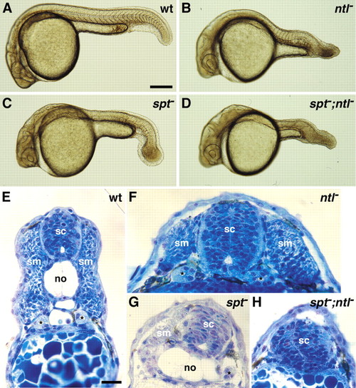
spt and ntl are required together for trunk and tail mesoderm development. (A-D) Live wild-type and mutant embryos at 24 hpf. Embryos doubly homozygous for spt and ntl mutations (D) have more severe posterior mesodermal deficiencies than ntl? (B) and spt? (C) embryos. Anterior development appears unperturbed. (E-H) Tranverse sections through trunk regions of wild-type (E) and mutant embryos (F-H) at 30 hpf show that (F) ntl? embryos lack a differentiated notochord, (G) spt? embryos have reduced muscle tissue, and (H) spt?;ntl? embryos lack all recognizable mesoderm, having only a spinal cord covered by skin. sc, spinal cord; no, notochord; sm, somitic muscle; *, pronephric tubule. Scale bars, 250 μm (A), 25 μm (E). |
|
Expression analyses reveal that mesoderm development, but not induction, is disupted in spt;ntl double mutant embryos. In situ hybridization and immunohistochemistry in wild-type (A,E,I,M), ntl? (B,F,J,N), spt? (C,G,K,O) and spt?;ntl? (D,H,L,P) embryos reveal synergistic and partially redundant interaction of spt and ntl during mesoderm formation. (A-D) ntl expression at mid-gastrulation (75% epiboly, 8 hpf) in wild-type and mutant embryos. (E-H) her1 expression during late gastrulation (95% epiboly, 9.5 hpf) in wild-type and mutant presomitic mesoderm. In I-L, pax2.1, krox20, and myoD transcripts are visualized in blue and Ntl protein is visualized in brown. In 10-somite stage (14 hpf) wild-type embryos (I), pax2.1 is expressed in the mid-hindbrain boundary, the otic placode and the developing pronephros (black asterisks), krox20 in hindbrain rhombomeres 3 and 5 (white asterisks), myoD in adaxial cells (arrowhead) and a subset of cells within each formed somite and the two most anterior forming somites, and Ntl in developing notochord cells. As discussed in the text, mesodermal gene expression is variably disrupted in ntl? and spt? embryos (J,K) and is abolished in spt?;ntl? embryos (L). (M-P) tropomyosin expression at 24 hpf. Embryos with intact yolks (A-H) are dorsal views, with anterior to the top. Dorsal (I-L) and lateral (M-P) views of deyolked embryos are shown with anterior to the left. Scale bars: 100 μm in A for A-D; E for E-H; I for I-L and M for M-P . EXPRESSION / LABELING:
PHENOTYPE:
|
|
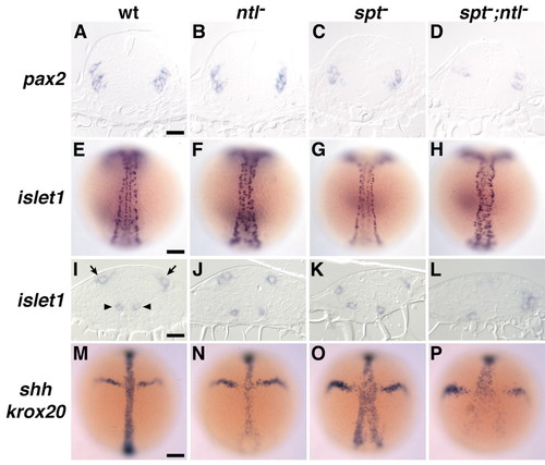
Neither spt nor ntl function is required for most dorsal-ventral spinal cord patterning. (A,E,I,M) Wild-type, (B,F,J,N) ntl?, (C,G,K,O) spt?, and (D,H,L,P) spt?;ntl? embryos. (A-D) pax2.1 interneuron expression at 24 hpf, (E-L) islet1 expression at the 8-10 somite stage (13-14 hpf); arrowheads indicate primary motoneurons and arrows indicate Rohon-Beard neurons. (M-P) sonic hedgehog (shh) and krox20 expression at the late gastrula stage (<9.5 hpf). Transverse sections (A-D; I-L) are approximately from midtrunk levels. Anterior is to the top. Scale bars: 25 μm (A-D and I-L), 100 μm (E-H and M-P). EXPRESSION / LABELING:
PHENOTYPE:
|
|
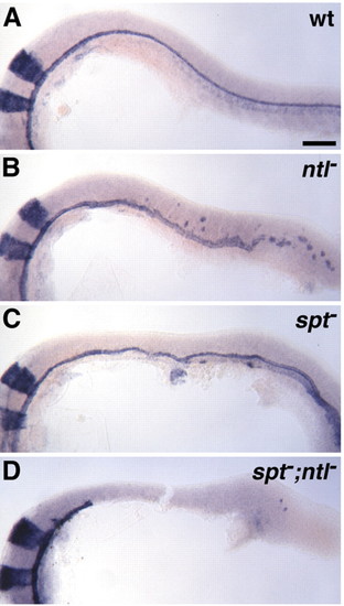
spt and ntl together are required for spinal cord medial floor plate formation. Expression of sonic hedgehog (shh) in ventral brain, medial floor plate (MFP), and posterior notochord and of krox20 in wild-type (A) and mutant (B-D) embryos at 24 hpf. Posterior MFP irregularities, such as broadening, are noted in ntl? (B) and spt? (C) embryos, but MFP is conspiciously missing in spt?;ntl? embryos (D), beginning approximately at the end of the hindbrain. Lateral views, with anterior to the left. Scale bar: 100 μm. |
|
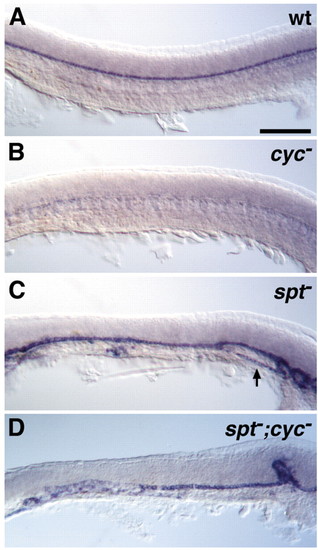
The nodal-related gene cyclops (cyc) is not required for medial floor plate development in the absence of spt function. sonic hedgehog (shh) expression in trunk medial floor plate (MFP) of wild-type (A) and mutant (B-D) embryos at 24 hpf. As noted already, spt? MFP is often broader and some MFP cells are located ventral to the notochord (arrow) (C). In contrast, MFP is completely absent in the trunk of cyc? embryos (B), but is significantly restored in spt?;cyc? embryos (D). Ventral brain shh expression in spt?;cyc? embryos is comparable to that in cyc? embryos (data not shown); thus, suppression of the cyc? MFP defect by the spt mutation is restricted to the trunk and tail. Lateral views, with anterior to the left. Scale bar: 100 μm. |
|
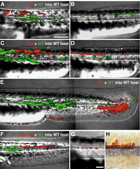
spt and ntl together are required cell-autonomously in mesodermal cells, but neither gene is required in medial floor plate cells. Genetic mosaic embryos were generated by isochronic transplantation of blastula cells from a rhodamine-labeled donor embryo derived from an intercross of two heterozygous spt;ntl carriers, together with blastula cells from a fluorescein-labeled wild-type donor, into the presumptive mesoderm region of a wild-type host embryo (see Materials and Methods for details). Shown are live embryos at approximately 36 hpf with rhodamine- and fluorescein-labeled cells visualized in red and green, respectively (A-G), and an embryo fixed at 22 hpf and processed to detect shh expression (blue) and a co-injected biotinylated-dextran lineage tracer (brown) (H). In A-F, the fluorescein-labeled cells are wildtype, and rhodamine-labeled cells are either wildtype (A,B), ntl? (C,D), spt? (E), or spt?;ntl? (F,G). Cells doubly homozygous for spt and ntl mutations never make mesoderm, but instead contribute to non-neural ectoderm, spinal cord, and MFP (F,G). Some genetic mosaics containing spt?;ntl? cells were fixed at approximately 22 hpf and processed to detect shh transcripts (blue) and the co-injected biotinylated-dextran lineage tracer (brown) to show that donor-derived spt?;ntl? floor plate cells express shh, a MFP marker (H). Scale bars: 100 μm (A-F), 25 μm (G,H). |
|
Excess ntl mutant floor plate cells originate from a fate map position corresponding to the wild-type notochord domain. Caged fluorescein fate mapping (see Materials and Methods and text) was used to determine fates of dorsal marginal cells of ntl? embryos. (A) The fluorophore was uncaged in the early gastrula in a small population of approximately 20 dorsal marginal cells of wild-type embryos and embryos depleted of ntl function (using ntl-MO). (B-E) Uncaged fluorescein label was detected at 24 hpf by fluorescence (B,C) and/or using anti-fluorescein antibody (D,E). Representative examples of deep layer cell (DEL) fates are shown. (B,D) In control wild-type embryos, the fluorophore was always detected in large populations of notochord cells, occasionally in hatching gland, and only rarely in floor plate. (C,E) In ntl-depleted embryos, the fluorophore was detected in large numbers of floor plate cells. See text for details. Scale bars: 50 μm, in A for A-C and in D for D,E. |

Unillustrated author statements PHENOTYPE:
|


