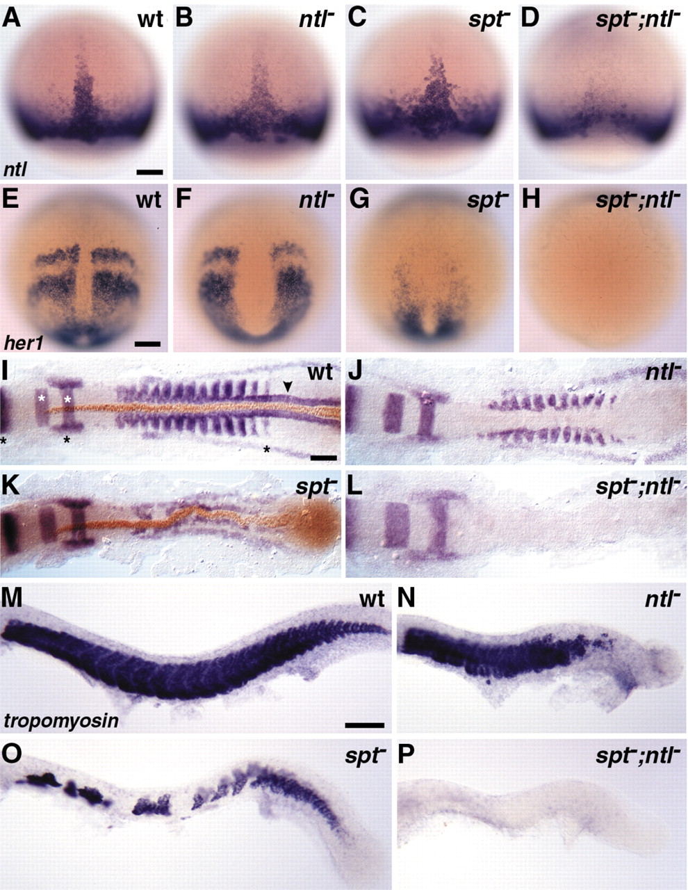
Fig. 3
Fig. 3 Expression analyses reveal that mesoderm development, but not induction, is disupted in spt;ntl double mutant embryos. In situ hybridization and immunohistochemistry in wild-type (A,E,I,M), ntl? (B,F,J,N), spt? (C,G,K,O) and spt?;ntl? (D,H,L,P) embryos reveal synergistic and partially redundant interaction of spt and ntl during mesoderm formation. (A-D) ntl expression at mid-gastrulation (75% epiboly, 8 hpf) in wild-type and mutant embryos. (E-H) her1 expression during late gastrulation (95% epiboly, 9.5 hpf) in wild-type and mutant presomitic mesoderm. In I-L, pax2.1, krox20, and myoD transcripts are visualized in blue and Ntl protein is visualized in brown. In 10-somite stage (14 hpf) wild-type embryos (I), pax2.1 is expressed in the mid-hindbrain boundary, the otic placode and the developing pronephros (black asterisks), krox20 in hindbrain rhombomeres 3 and 5 (white asterisks), myoD in adaxial cells (arrowhead) and a subset of cells within each formed somite and the two most anterior forming somites, and Ntl in developing notochord cells. As discussed in the text, mesodermal gene expression is variably disrupted in ntl? and spt? embryos (J,K) and is abolished in spt?;ntl? embryos (L). (M-P) tropomyosin expression at 24 hpf. Embryos with intact yolks (A-H) are dorsal views, with anterior to the top. Dorsal (I-L) and lateral (M-P) views of deyolked embryos are shown with anterior to the left. Scale bars: 100 μm in A for A-D; E for E-H; I for I-L and M for M-P .

