
|
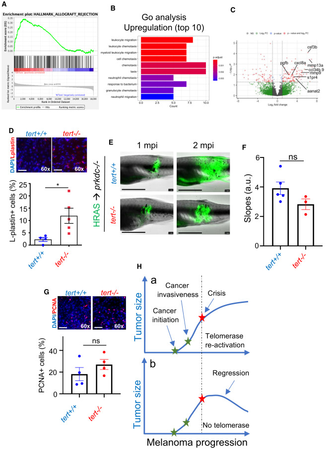
Fig. 5 The immune system restrains growth of telomerase-deficient melanoma Telomerase-deficient tumors are strongly immunogenic. (A) Allograft rejection enrichment plot. (B) Gene Ontology (GO) enrichment analysis for upregulated genes of late-stage melanoma of tert?/? fish compared to tert+/+. (C) Volcano plot of differentially expressed genes of late-stage melanoma of tert?/? fish compared to tert+/+, highlighting genes included in leukocyte migration pathway from the GO analysis. (D) Immunofluorescence images and quantifications of immune cell infiltrates (L-plastin) of late-stage melanoma. Late-stage tert?/? melanoma is able to proliferate and grow in immunocompromised fish (n ? 4; unpaired t test). Scale bars: 20 ?m. (E) Images of immunocompromised zebrafish (prkdc?/?) engrafted with primary melanoma cells derived from 9-month-old zebrafish at 1 and 2 months post-injection (mpi). Scale bars: 0.5 cm. (F) Progression slope of tumor area calculated by a linear regression of two time points (1 and 2 mpi) (n ? 4; unpaired t test). (G) Cell proliferation of tumor cells quantified using PCNA immunofluorescence (n ? 4; unpaired t test). Scale bars: 20 ?m. (H) We propose that melanoma can initiate and progress without telomerase activation. However, once telomeres become critically short, cancer cells must activate telomerase to sustain proliferation at later stages (a). If they fail to do so, tumor growth stagnates, leading to tumor regression (b). Error bars represent � SEM; each dot represents an individual tumor; ?p ? 0.05. ns, not significant.