Image
Figure Caption

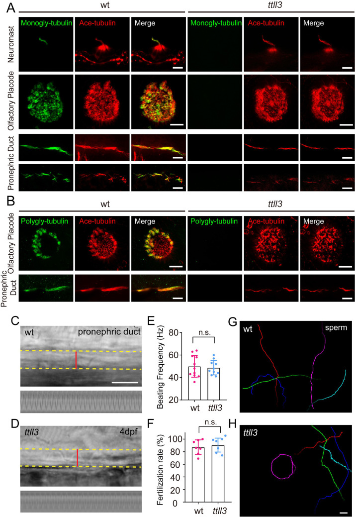
Figure 4—figure supplement 2—source data 1. Loss of tubulin glycylation in ttll3 mutants.

(A) Immunostaining with anti-monoglycylated tubulin antibody in 4 dpf ttll3 mutants showing complete absence of monoglycylation modification in cilia of neuromast, olfactory placode and pronephric duct. (B) Immunostaining with anti-polyglycylated tubulin antibody revealed complete elimination of polyglycylation modification in multicilia of olfactory placode and pronephric duct. Red channel shows cilia visualized with anti-acetylated α-tubulin antibody. (C, D) Snapshots of high-speed video showing cilia in pronephric duct of 5 dpf wild type (C) and ttll3 (D) mutant. Bottom images show kymographs of cilia beating. The yellow dashed line indicates the boundary of pronephric duct, and the red line represents the path used to generate the kymograph. (E) Beating frequency of cilia in pronephric duct of wild type and ttll3 mutant (wt, n=11; ttll3, n=10, Unpaired two-sided Student’s t-test). (F) Statistical results showing fertilization rate of wild type and ttll3 mutant (n=7, Unpaired two-sided Student’s t-test). (G, H) Sperm movement trajectory of wild type (G) and ttll3 (H) mutant within 2 s. Scale bar = 5 μm in panel A and B, and 10 μm in panel C, D, G and H.

Acknowledgments
This image is the copyrighted work of the attributed author or publisher, and ZFIN has permission only to display this image to its users. Additional permissions should be obtained from the applicable author or publisher of the image. Full text @ Elife