IMAGE

Figure 1.

ID
ZDB-IMAGE-241218-35
Source
Figures for Sun et al., 2024
Image
Figure Caption

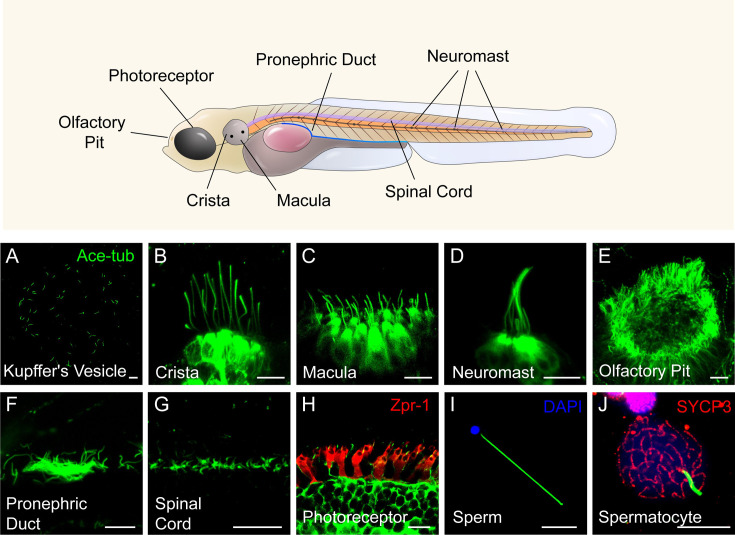
Figure 1. Diverse type of cilia are present in zebrafish.

(A) Cilia in kupffer’s vesicle (KV) of a 10-somite stage zebrafish larvae. (B–H) Confocal images showing cilia in different type of cells at 4 dpf as indicated. The position of these cells were indicated in the top diagram. (I–J) Confocal images showing cilia in the sperm (flagellum) or spermatocyte of adult zebrafish. All the cilia were visualized with anti-acetyleated tubulin. The photoreceptor double cones were stained with zpr-1 antibody in panel H, and nuclei were stained with DAPI in panel I. Immunostaining with anti-SYCP3 labelled the synaptonemal complexes of primary spermatocytes in panel J. Scale bar = 10 μm.

Acknowledgments
This image is the copyrighted work of the attributed author or publisher, and ZFIN has permission only to display this image to its users. Additional permissions should be obtained from the applicable author or publisher of the image. Full text @ Elife