IMAGE

Fig. 1

ID
ZDB-IMAGE-241018-1
Source
Figures for Li et al., 2024
Image
Figure Caption

Fig. 1

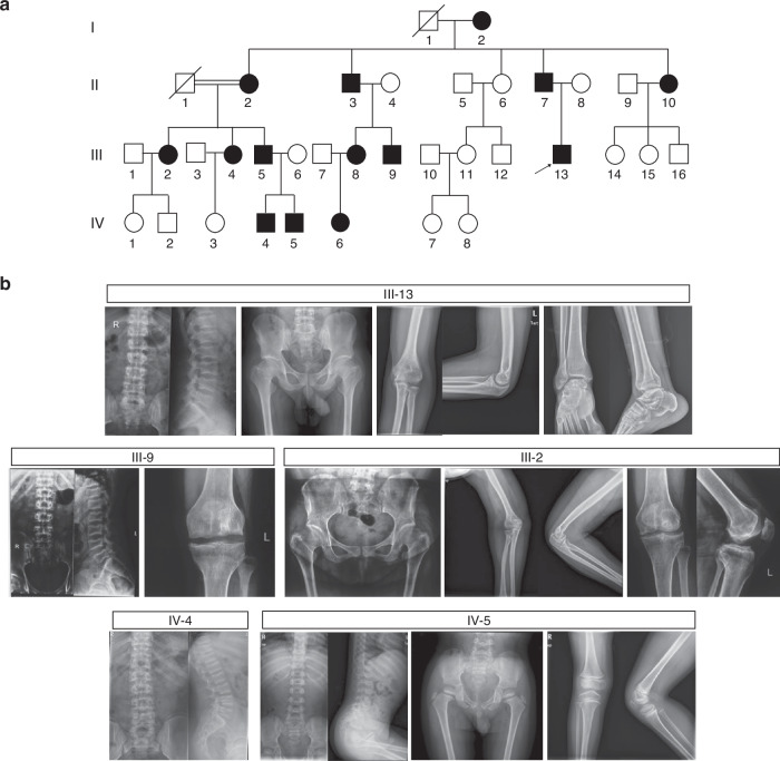
Pedigree and radiographical features of a large Chinese family with spondyloepimetaphyseal dysplasia (SEMD). a Pedigree of the family with SEMD. Squares and circles indicate males and females, respectively. Black symbols present affected individuals, and slashes deceased individuals. Arrow indicates the proband. b Radiographic abnormalities of subjects with SEMD. III-13: individual at age 25.7 years. Spine lateral shows platyspondyly and biconcave deformities of the central-posterior portion of vertebral bodies, with irregular sclerotic end plates. Pelvis shows mildly narrow iliac wings with irregular acetabulum and femoral head. Short and broad femoral necks are noted. Elbow and ankle joint show irregular proximal humerus and distal tibiae and fibulae, as well as hyperostosis. The joint spaces are preserved. III-9: individual at age 28.9 years. Platyspondyly with irregular sclerotic end plates, and biconcave vertebral bodies are noted. Knee joint shows mildly decreased joint spaces, irregular articular surface, osteophyte formation, together with sclerotic and cystic changes. III-2: individual at age 44.6 years. Femoral necks are short and broad, with irregular acetabuli and femoral head. Degenerative changes, including narrow joint cavity, hyperostosis, subchondral cysts and bone sclerosis are noted in hip, elbow and knee joints. IV-4: individual at age 15.1 years. Spine lateral shows flat vertebral bodies, with irregular epiphyses and mild scoliosis. IV-5: individual at age 4.6 years. Distinctive bullet-shaped appearance vertebral bodies and irregular epiphyses are noted. Pelvis and knee joint show irregular epiphyses and metaphyses of the long bones, together with acetabular abnormalities

Acknowledgments
This image is the copyrighted work of the attributed author or publisher, and ZFIN has permission only to display this image to its users. Additional permissions should be obtained from the applicable author or publisher of the image. Full text @ Bone Res