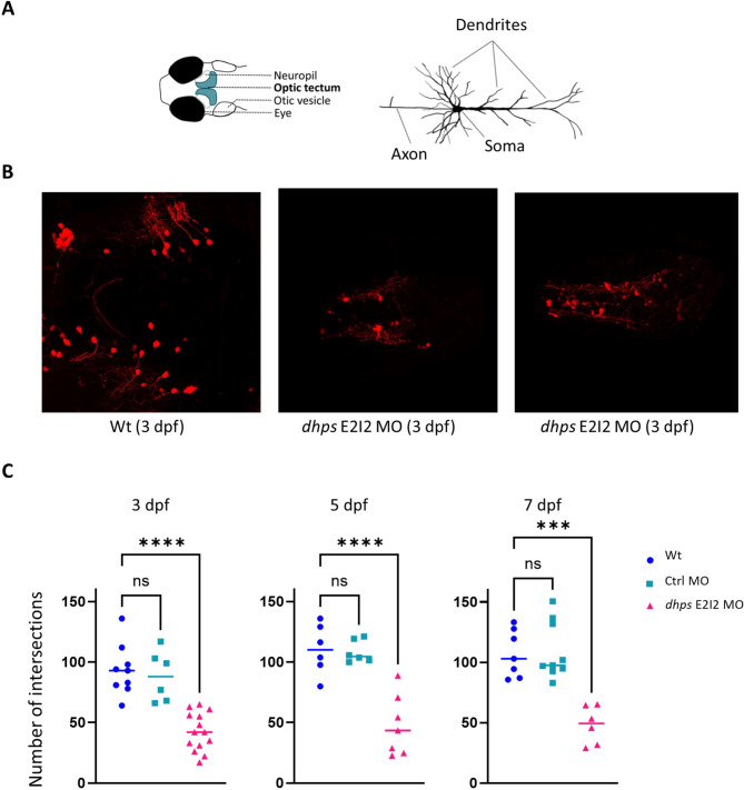
IMAGE

Fig. 5

ID
ZDB-IMAGE-240930-121
Source
Figures for Shojaeinia et al., 2024
Image
Figure Caption

Fig. 5

GABAergic neuronal dendritic arborization. (A) Larval head with location of optic tectum (blue) and neuropil (light yellow) where that GABAergic neurons project their arbors. (B) Confocal microscopy revealed that dhps knockdown larvae have less complex dendritic arborization in the GABAergic neurons. (C) GABAergic neuronal arbors were quantified in zebrafish larvae at three different ages (3, 5, and 7 dpf). At all three developmental ages, via one way ANOVA there was a significant reduction in the number of neuronal arbors in the dhps knockdown larvae (dhps E2I2 MO) versus both wild-type control larvae (Wt) and control MO-injected larvae (Crtl MO) (see also Table 1 below)

Acknowledgments
This image is the copyrighted work of the attributed author or publisher, and ZFIN has permission only to display this image to its users. Additional permissions should be obtained from the applicable author or publisher of the image. Full text @ Mol. Brain