
|
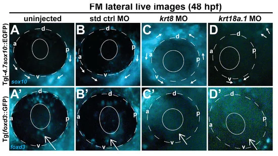
Fig. 4 Krt8/Krt18a.1 MO knockdown disrupted ocular neural crest cell migration dynamics during early eye development. Lateral live imaging at 48 hpf shows the effects of Krt8 and Krt18a.1 knockdown on GFP reporter expression in Tg(foxd3::EGFP) and Tg(-4.7sox10::EGFP) zebrafish embryos injected at the single-cell stage with antisense MOs targeting krt8 and krt18a.1. (A,A′) uninjected, (B,B′) standard control (std ctrl) MO-injected, (C,C′) krt8 MO-injected, and (D,D′) krt18a.1 MO-injected. The migration of GFP-positive neural crest cells into the ocular (open arrow) and periocular (solid arrows) regions was markedly disrupted in response to Krt8/Krt18a.1 MO knockdown compared with that in uninjected and control MO-injected embryos. The solid and dashed circles highlight the lens and retinal pigment epithelium, respectively, of the zebrafish eye. d, dorsal; v, ventral; p, posterior; a, anterior.