
Image
|
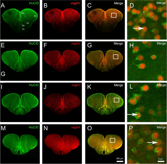
Figure Caption
Fig. 10 vegf receptor expression is observed in most neurons within the telencephalon. A-P vegfr1, vegfr2, vegfr3, and vegfr4 in situ hybridization (red) followed by HuC/D immunohistochemistry (green) in the telencephalon. D, H, L and P High magnification views of the respective white squares in the telencephalon showing that most vegfr-positive cells correspond to HuC/D-positive neurons. Bars: 200 �m (A-C, E?G, I-K, M?O) and 6 �m (D, H, L and P)
Acknowledgments
This image is the copyrighted work of the attributed author or publisher, and
ZFIN has permission only to display this image to its users.
Additional permissions should be obtained from the applicable author or publisher of the image.
Full text @ Neural Dev.