Image
Figure Caption
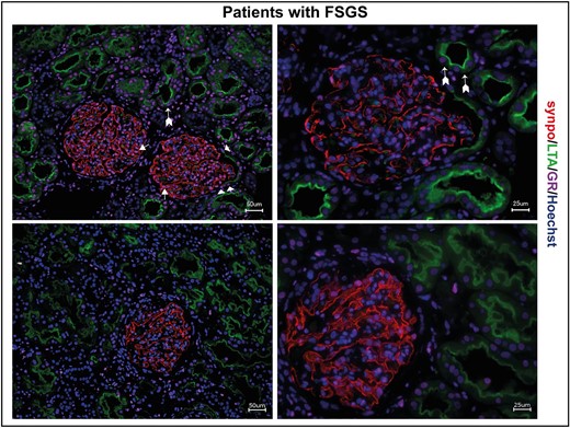
Fig. 1 GR expression in human MCD/FSGS. Immunofluorescence staining of human kidney biopsies of two patients with FSGS. GR expression (magenta) is visible in all glomerular cells, including podocytes (synaptopodin; red, arrow) and parietal epithelial cells (arrowheads), but also in proximal tubule cells (LTA; green, arrows with tails). Scale bars 25 ?m and 50 ?m.
Acknowledgments
This image is the copyrighted work of the attributed author or publisher, and
ZFIN has permission only to display this image to its users.
Additional permissions should be obtained from the applicable author or publisher of the image.
Full text @ Nephrol. Dial. Transplant.

