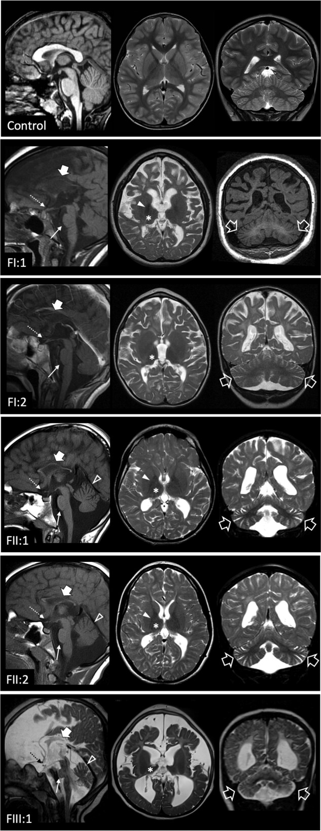
Fig. 2 Neuroimaging findings. Brain MRI studies of a control subject performed at 9 years of age for comparison, and of the patients performed at 9 years (Patient FI:1), 7.7 years (Patient FI:2), 13 years Patient FII:1), 12 years (Patient FII:2) and 3 years of age (Patient FIII:1). In the patients, sagittal T1- or T2-weighted (left), axial T2-weighted (middle) and coronal T1 or T2-weighted images (right) reveal mild-to-severe cerebral atrophy with reduced white matter volume and enlarged subarachnoid spaces in all cases. There is diffuse T2 hyperintensity of the cerebral white matter in all subjects, with relative sparing of the internal capsules and subcortical U-fibres in Patients FII:1 and FII:2, in keeping with hypomyelination. Focal signal alterations are associated at the level of the fronto-parietal white matter. The corpus callosum is thin in all patients (thick arrows). The thalami are very small and slightly T2 hypointense in all cases (asterisks). The globi pallidi are small and darker on T2-weighted images in Patients FI:1, FII:1 and FII:2 (full arrowheads). There is mild pontine hypoplasia (thin arrows) and marked optic nerve and chiasm atrophy (thin dashed arrows) in all subjects. Mild-to-moderate cerebellar atrophy with prevalent enlargement of the hemispheric subarachnoid spaces is present in all subjects (empty arrows), while involvement of the superior vermis is visible only in Patients FII:1, FII:2 and FIII:1 (empty arrowheads).
Image
Figure Caption
Acknowledgments
This image is the copyrighted work of the attributed author or publisher, and
ZFIN has permission only to display this image to its users.
Additional permissions should be obtained from the applicable author or publisher of the image.
Full text @ Brain

