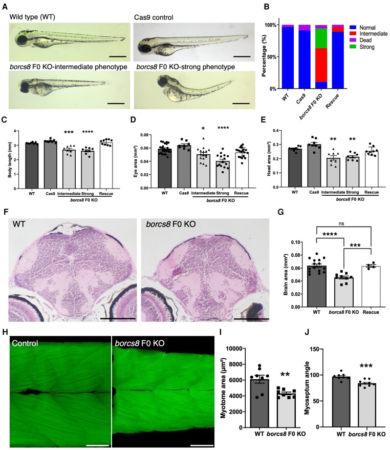
Fig. 6 Zebrafish borcs8 F0 knockout larvae exhibit developmental defects. (A) Morphology of zebrafish wild-type (WT), Cas9 control and borcs8 F0 knockout (KO) larvae at 3 days post-fertilization (dpf). Scale bars = 1 mm. (B) Frequency of phenotypes observed for wild-type, Cas9 control, borcs8 F0 KO and human BORCS8 mRNA rescue (Rescue) larvae (N = 4, n = 32?72). (C?E) Body length (C), head size (D) and eye size (E) of wild-type, Cas9 control, borcs8 F0 KO and Rescue larvae at 3 dpf (N = 3, n = 8?9 for head size and body length, n = 16?18 for eye size). (F) Haematoxylin and eosin staining of paraffin-embedded brain sections from the midbrain of 5 dpf wild-type and borcs8 F0 KO larvae. Scale bar = 100 ?m. (G) Quantification of brain size of borcs8 F0 KO larvae (N = 9) relative to wild-type (N = 15) and Rescue (N = 4) at 5 dpf. (H) Phalloidin staining of muscles in borcs8 F0 KO and wild-type larvae at 3 dpf. (I and J) Comparisons of dorsal or ventral myotome area (I) and myoseptum angle (J) between wild-type (N = 3, n = 8) and borcs8 F0 KO larvae (N = 3, n = 8?9). All data are represented as the mean ± standard error of the mean (SEM). Statistical significance was calculated by one-way ANOVA followed by Tukey?s multiple comparisons tests, or Student?s t-test. *P < 0.05; **P < 0.01; ***P < 0.001; ****P < 0.0001. N = replicates; n = sample size; ns = not significant.
Image
Figure Caption
Figure Data
Acknowledgments
This image is the copyrighted work of the attributed author or publisher, and
ZFIN has permission only to display this image to its users.
Additional permissions should be obtained from the applicable author or publisher of the image.
Full text @ Brain

