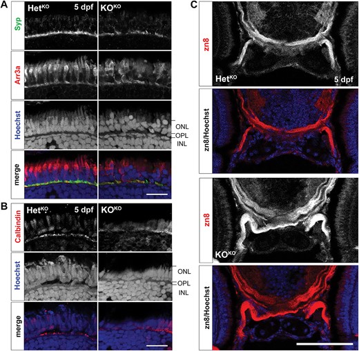
Fig. 9 Retinal and axon guidance phenotypes are found at 5 dpf in pomt1 KOKO larvae. (A) Discontinuities in synaptophysin (Syp) staining are found in the outer plexiform layer (OPL) of pomt1 KOKO retinae. Red/green double cone marker arrestin 3a (Arr3a) again shows disorganized outer segments and patchy staining and altered shape of photoreceptors pedicles. Nuclear staining with Hoechst shows disorganization disruption in the photoreceptor nuclei in the outer nuclear layer (ONL) and in horizontal cell nuclei at the top of the inner nuclear layer (INL). Scale bar: 20 μm. (B) Horizontal cell dendrites extending in the OPL are visualized using anti-calbindin staining and are disrupted in the KOKO retina. Scale bar: 20 μm (C) The optic nerve was labeled using the zn8 antibody that marks retinal ganglion cell axons showing axon defasciculation after optic chiasm crossing. Imaged including the full head are shown in Supplementary Fig. 8B. Scale bar: 50 μm. Alt-text. This figure shows that the same photoreceptor synapse loss phenotypes found at 30 dpf in Fig. 4 are already present at 5 dpf in pomt1 knock-out fish obtained from knock-out mothers. In addition, there is defasciculation of the optic tract indicating axon guidance deficits.
Image
Figure Caption
Figure Data
Acknowledgments
This image is the copyrighted work of the attributed author or publisher, and
ZFIN has permission only to display this image to its users.
Additional permissions should be obtained from the applicable author or publisher of the image.
Full text @ Hum. Mol. Genet.

