
Image
|
Figure Caption
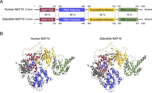
Fig. 1 Comparative analysis of human and zebrafish NAT10 proteins. (A) Domain-wise percent sequence identity between human and zebrafish NAT10 proteins, highlighting their conserved regions. (B) Predicted three-dimensional (3D) structures of human and zebrafish NAT10 proteins using AlphaFold, with distinct domains, color-coded: DUF1726 (purple), RNA Helicase (blue), N-acetyltransferase (yellow), and RNA binding (green).
Acknowledgments
This image is the copyrighted work of the attributed author or publisher, and
ZFIN has permission only to display this image to its users.
Additional permissions should be obtained from the applicable author or publisher of the image.
Full text @ Invest. Ophthalmol. Vis. Sci.