IMAGE

Fig. 6

ID
ZDB-IMAGE-240323-6
Source
Figures for Ma et al., 2024
Image
Figure Caption

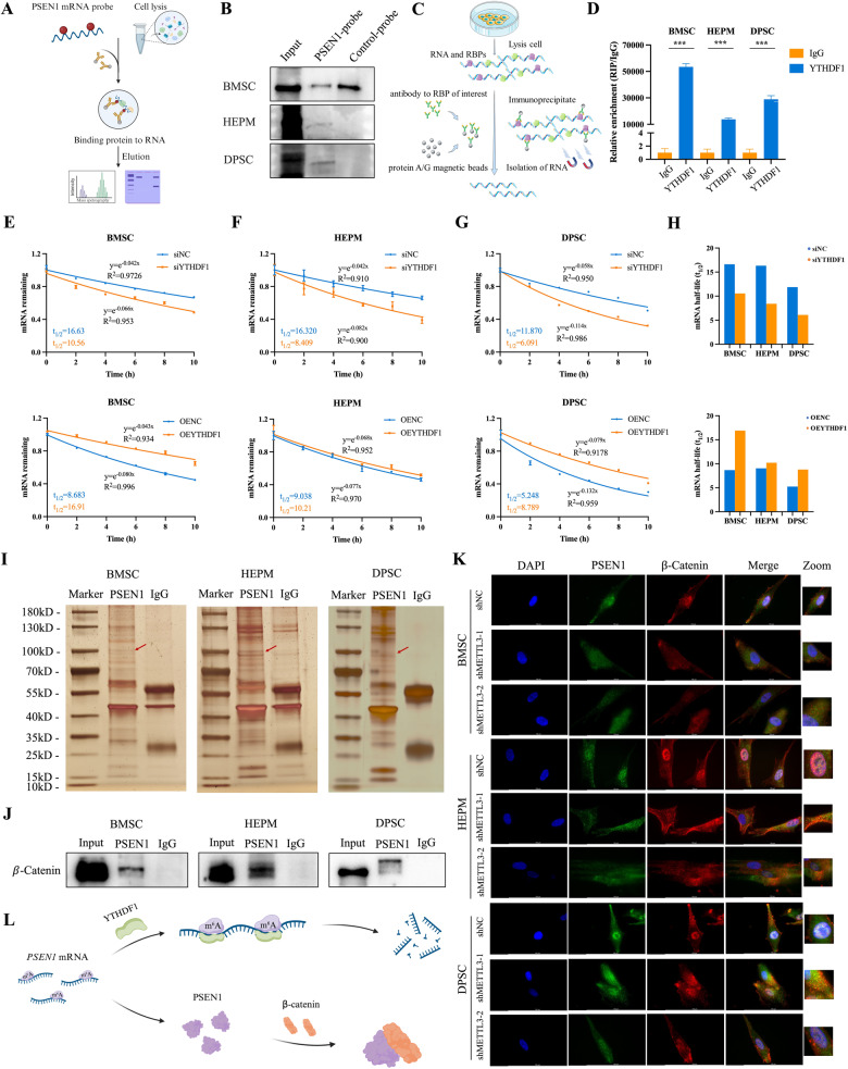
Fig. 6 PSEN1 is specifically recognized by YTHDF1 and directly interacts with β-catenin.

A Schematic of the design for the RNA pull-down assay. B Immunoblotting of YTHDF1 after the RNA pull-down assay with cell lysate, biotinylated-PSEN1, and biotinylated-control in the cells. C Schematic of the design for the RNA immunoprecipitation (RIP) assay. D RIP assay to determine the enrichment of PSEN1 in cells incubated with anti-YTHDF1 antibody. EH Cells were transiently transfected with control, siYTHDF1, empty vector, or OEYTHDF1, respectively. The half-life (t1/2) of the PSEN1 mRNA was measured. I Silver staining revealed PSEN1-bound proteins in BMSCs, HEPM cells and DPSCs. J Interaction between β-catenin and PSEN1 determined by co-IP followed by western blot analysis. K Representative image showing the enrichment of PSEN1 and β-catenin after METTL3 knockdown by immunostaining analyses in BMSCs, HEPM cells, and DPSCs. L Schematic diagram showing the mechanism of PSEN1. Results were presented as mean ± SD of three independent experiments. ***P < 0.001 indicates a significant difference between the groups.

Acknowledgments
This image is the copyrighted work of the attributed author or publisher, and ZFIN has permission only to display this image to its users. Additional permissions should be obtained from the applicable author or publisher of the image. Full text @ Cell Death Dis.