IMAGE

Figure 2

ID
ZDB-IMAGE-231127-29
Source
Figures for Hall et al., 2023
Image
Figure Caption

Figure 2

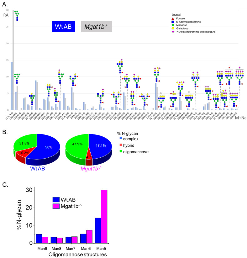
Comparison of LC-ESI-MS relative intensities of the permethylated N-glycans derived from Wt AB and the Mgat1b/ mutant line. LC-ESI-MS intensities of released N-glycans from Wt AB (blue bars) and Mgat1b/ (grey bars) (A). All molecular ions are present in sodiated form ([M + Na]+). N-glycan numbers correspond to Figures S1 and S2, and Tables S2 and S3. Breakdown of the percent of various detected N-glycan types (B) and types of oligomannose structures (C) identified by the ESI-MS spectra.

Figure Data
Acknowledgments
This image is the copyrighted work of the attributed author or publisher, and ZFIN has permission only to display this image to its users. Additional permissions should be obtained from the applicable author or publisher of the image. Full text @ Curr. Iss. Mol. Biol.