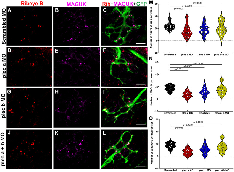
Fig. 7 Plectin knockdown results in abnormal hair cell synapses. A-L: Representative images of 3dpf Tg(NeuroD-GFP) neuromasts from scrambled (A-C), plectin a (D-F), plectin b (G-I), plectin a + b (J-L) morphants. Samples were immunostained for the pre-synaptic marker, ribeye B (red), the post-synaptic marker, MAGUK (magenta), and the afferent neuronal marker, GFP (green). Scale bar: 5 ?m. M-O: Violin plots comparing the number of pre-synaptic puncta (M), post-synaptic puncta (N) and ribbon synapses (O) in the different groups. Each dot represents the number of puncta per neuromast, with at least two neuromasts inspected per fish, and between 6 and 10 fish inspected. One-way ANOVA followed by Dunnett T3 multiple comparisons test.
Reprinted from Hearing Research, 436, Zhang, T., Xu, Z., Zheng, D., Wang, X., He, J., Zhang, L., Zallocchi, M., Novel biallelic variants in the PLEC gene are associated with severe hearing loss, 108831108831, Copyright (2023) with permission from Elsevier. Full text @ Hear. Res.

