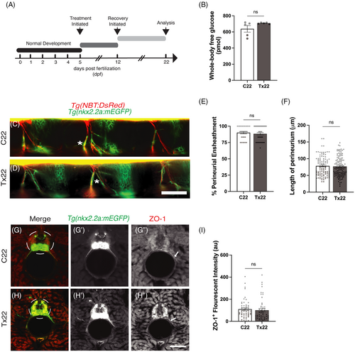
Fig. 2 Regeneration of the perineurial structure ensheathing the motor nerves following recovery from acute hyperglycemia. (A) A schematic outlining the treatment and recovery schedule. (B) Following the schedule of treatment described in (A), there was no significant (ns) difference in whole-body glucose levels of Tx22 fish compared to C22 fish, P = .1284. (C and D) Lateral view of C22 and Tx22 Tg(nkx2.2a:mEGFP);Tg(NBT:DsRed) zebrafish. Following the recovery period, perineurial glia (green) were associated with peripheral motor axons (red) in both C22 (C, asterisk) and Tx22 (D, asterisk) fish. (E) The percentage of perineurial-ensheathed motor axons did not differ between Tx22 and C22 fish following recovery, P = .4730. (F) Similarly, no difference in the length of perineurial-ensheathment was observed between C22 and Tx22 fish following the recovery period, P = .6890. (G and H) Representative images of C22 and Tx22 Tg(nkx2.2a:mEGFP) zebrafish antibody labeled against ZO-1 (red). ZO-1+ tight junction proteins were continuous along the length of the perineurium in C22 fish (G?, arrow). ZO-1+ tight junction proteins also appeared to be continuous in Tx22 fish (H?, arrow). (I) ZO-1+ fluorescent intensity (standardized to nkx2.2a) was not significantly different between Tx22 and C22 fish, P = .8400 (au, arbitrary units). All values are means ± SEM. Scale bars = 50 ?m.
Image
Figure Caption
Acknowledgments
This image is the copyrighted work of the attributed author or publisher, and
ZFIN has permission only to display this image to its users.
Additional permissions should be obtained from the applicable author or publisher of the image.
Full text @ Dev. Dyn.

