
|
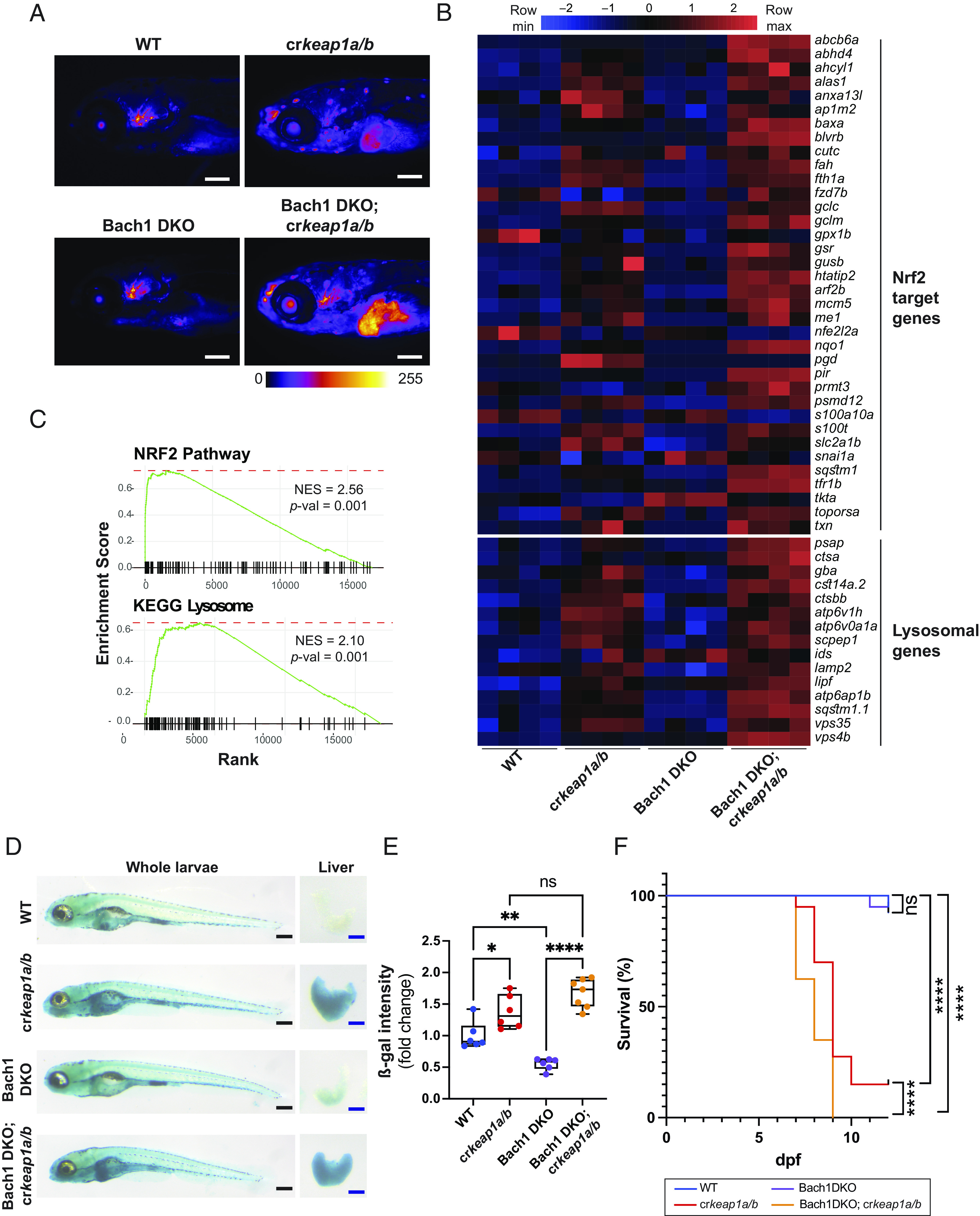
Fig. 4.
Bach1-mediated repression of Nrf2 modulates lysosomal biogenesis. (A) Representative whole-mount fluorescent images of WT, crkeap1a/b, Bach1 DKO, and Bach1 DKO; crkeap1a/b zebrafish on a gstp1:EGFP background at 7 dpf. Fluorescent images are pseudocolored using the Fire LUT. Scale bars represent 200 µm. (B) Heatmap of Nrf2 target genes and lysosomal genes in WT, crkeap1a/b, Bach1 DKO, and Bach1 DKO; crkeap1a/b zebrafish at 7 dpf as determined by RNA-Seq analysis, n = 4 pools of 10 larvae. (C) GSEA plots derived from RNA-Seq analysis of Bach1 DKO; crkeap1a/b versus Bach1 DKO zebrafish at 7 dpf demonstrating the Nrf2 pathway signature and KEGG lysosomal signature. (D) Representative images of ß-gal-stained whole-mount (Left) and dissected larval livers (Right) from WT, crkeap1a/b, Bach1 DKO, and Bach1 DKO; crkeap1a/b zebrafish at 7 dpf. Black scale bars represent 350 µm, blue scale bars represent 100 µm. (E) Quantification of ß-gal intensity in dissected larval livers represented in D. Data are shown as mean and interquartile range, n = 6 to 7. (F) Kaplan–Meier survival plot of WT, crkeap1a/b, Bach1 DKO, and Bach1 DKO; crkeap1a/b zebrafish, n = 40. For all experiments, *P < 0.05, **P < 0.01, ****P < 0.0001.