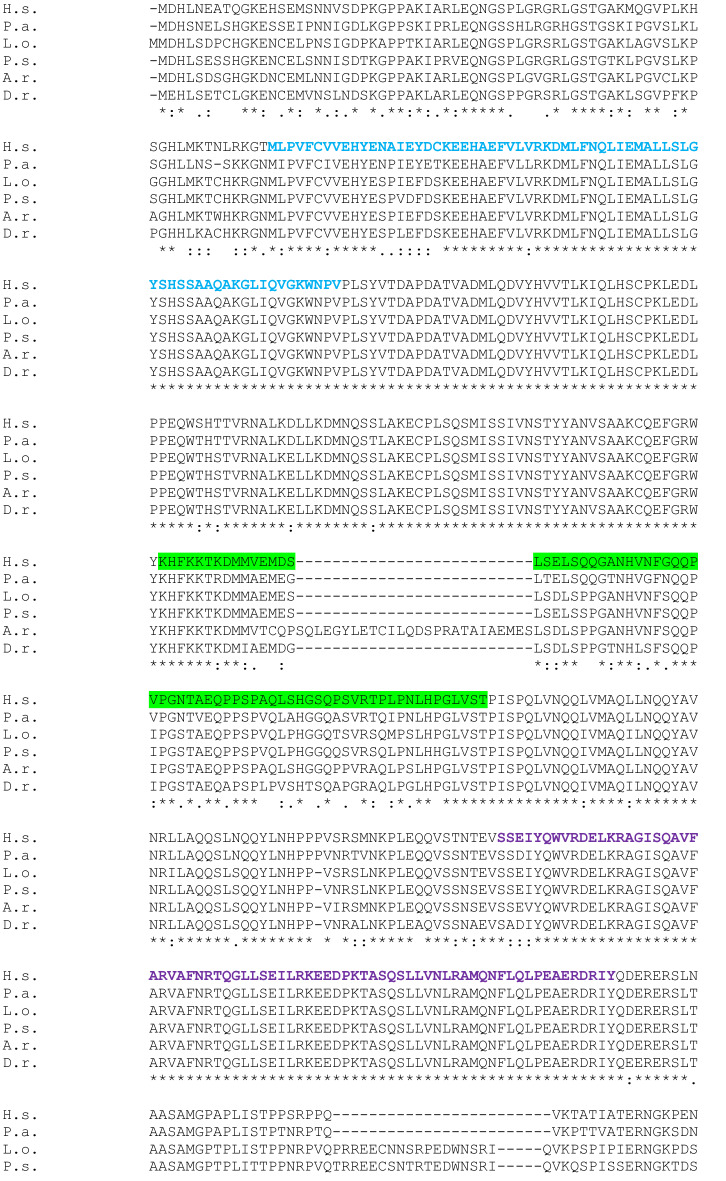
Fig. 2
Full-length SATB1 protein sequence alignment in Homo sapiens (H.s) as a query, and Protopterus annectens (P.a), Lepisosteus oculatus (L.o), Danio rerio (D.r), Acipenser ruthenus (A.r) and Polypterus senegalus (P.s) using CLUSTALW. Identical amino acids are shown with an asterisk (*). The colon (:) and the dot (.) indicate a conserved substitution, and a semi-conserved substitution respectively. The SATB domain is shown in blue, the CUT domains are shown in purple and the homeodomain in yellow. The epitope sequence recognized by the Satb1/2 antibody (Abcam. Catalog reference ab51502) is underlined in red and the epitope sequence recognized by the Satb1 antibody (Santa Cruz Biotechnology. Catalog reference sc-376096) is underlined in green

