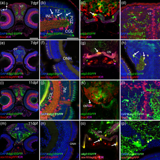
FIGURE 4
olig2:EGFP cells are differentiated from 7 dpf. In the retina at 7 dpf, olig2:EGFP cells are restricted to the PGZ and the TZ (a, b). But only the cells in the TZ colocalize with Sox2 (arrows in b). Sox2 that do not show olig2:EGFP are abundant in the proliferative areas of the brain (c, d). olig2:EGFP occasionally colocalize with Sox2 in the optic nerve chiasm at 7 dpf (arrow in c). At 7 dpf olig2:EGFP/sox10:tagRFP cells were found in the ON chiasm (e, arrow in g) and in other parts of the brain (arrows in h). Differentiated part of the retina was empty of olig2:EGFP/sox10:tagRFP cells (f). At 11 dpf, Sox2 keeps its pattern in the retina (i, j) and in the ON (arrow k). In the OT, Sox2 cells are restricted to the ventricular area (l). olig2:EGFP/sox10:tagRFP cells are abundant in the ON with obvious projections (m, arrows in o) and there are none in the retina (n). Oligodendrocytes in the OT are olig2:EGFP/sox10:tagRFP (p). Calretinin (CR) is used to label ganglionar cells and ON. D: dorsal; GCL: ganglion cell layer; INL: inner nuclear layer; L: lateral; ONH: optic nerve head; ON; optic nerve; OT: optic tectum; PGZ: proliferative germinal zone; VZ: ventricular zone. Scale bar in a, e, i, m: 100 μm; in b, c, d, f, g, h, j, k, l, n, o, p: 50 μm

