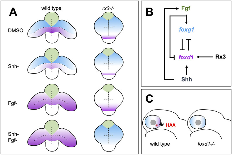
Fig. 4. Summary of roles for Rx3, Fgfs and Shh in establishment of nasal and temporal character in the developing eye primordium. (A) Schematic representation of wild-type and rx3?/? embryos depicting the changes in foxg1 (blue) and foxd1 (magenta) expression upon abrogation of Shh and Fgf activities, singly or in combination. (B) Diagram detailing the proposed regulatory network controlling NT patterning. Shh promotes foxd1 expression in the ventral (future temporal) half of the optic primordium. Fgfs induce foxg1 in the dorsal (future nasal) optic primordium and repress foxd1 expression, contributing to its confinement ventrally. Cross-repression between Foxg1 and Foxd1 at the border between the dorsal and ventral halves of the eye primordium subsequently refines the naso-temporal subdivision. Although induction of shh and fgf8 expression occurs independently, fgf8 expression is lower in the absence of Shh signalling (Hernández-Bejarano et al., 2015), suggesting Hh signalling initially promotes Fgf signalling. Rx3 induces foxd1 expression and provides competence to optic vesicle cells to enhance foxd1 expression in prospective temporal retina in response to Shh signalling. (C) Schematic representation of a differentiated optic cup with nasal (blue) and temporal (magenta) character, and the HAA (red) highlighted. The nasal character is expanded and HAA and temporal character are absent in foxd1?/? mutants (right) when compared with the wild type (left).
Image
Figure Caption
Acknowledgments
This image is the copyrighted work of the attributed author or publisher, and
ZFIN has permission only to display this image to its users.
Additional permissions should be obtained from the applicable author or publisher of the image.
Full text @ Development

