IMAGE

Fig. 5

ID
ZDB-IMAGE-220901-18
Source
Figures for Liu et al., 2022
Image
Figure Caption

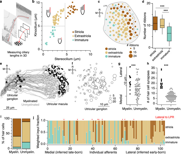
Fig. 5 The utricular afferent ganglion is organized in the mediolateral axis by developmental sequence and temporal dynamics.

a Representation of kinocilium reconstruction across successive images to obtain the total length. The neighboring tallest stereocilium was also reconstructed (not schematized). b Plot of length of the kinocilium vs. the tallest stereocilium for all 91 hair cells. Striolar hair cells are identified by their long stereocilia and low K/S ratios, whereas extrastriolar hair cells have higher K/S ratios. Hair cells characterized as immature have short kinocilia and stereocilia. c Horizontal projection of the utricular macula, showing number of synaptic ribbons in each hair cell. Circle diameter reflects synaptic ribbon count; hair cells with larger numbers of ribbons tend to be located more centrally. See also Supplementary Fig. 1d Quantification of synaptic ribbon counts across hair cell categories. Box plot represents medians ± 25%ile; whiskers indicate 10-90%iles. Immature hair cells form fewer ribbon synapses than striolar or extrastriolar hair cells. N = 23 striolar, 52 extrastriolar, and 16 immature hair cells. Significance was tested with ANOVA (p = 4.8 × 10−5) and then pairwise with Wilcoxon–Mann–Whitney (striola—extrastriolar, p = 0.60; striolar—immature, p = 4.3 × 10−4; extrastriolar—immature, p = 1.3 × 10−4). e Horizontal projection of all utricular afferents, colorized by whether they are myelinated or not. f Horizontal projection map of all utricular ganglion somata by position; circle diameter reflects the number of hair cell ribbon synaptic inputs that each one receives. g Myelinated afferent somata are located more laterally in the utricular ganglion. Each dot represents one afferent soma. Wilcoxon–Mann–Whitney, p = 2.9 × 10−4. h Myelinated afferents are contacted by significantly more hair cell ribbons than unmyelinated afferents. Wilcoxon–Mann–Whitney, p = 1.8 × 10−10. See also Supplementary Fig. 3c. i Myelinated afferents receive the majority of their input from striolar hair cells, whereas unmyelinated afferents receive most of their input from extrastriolar and developing hair cells. The distributions are significantly different (chi-squared test, p < 1 × 10−10). See also Supplementary Fig 3d, e. j The weighted fraction of inputs each utricular afferent receives from the different hair cell classes. Afferents are ordered based on soma position from medial to lateral. Red dots identify afferents receiving input from hair cells lateral to the LPR (contralateral tilt sensitive). Laterally positioned afferents typically have predominantly striolar inputs, whereas medially positioned afferents have predominantly extrastriolar or immature inputs.

Acknowledgments
This image is the copyrighted work of the attributed author or publisher, and ZFIN has permission only to display this image to its users. Additional permissions should be obtained from the applicable author or publisher of the image. Full text @ Nat. Commun.