IMAGE

Fig. 3

ID
ZDB-IMAGE-220901-16
Source
Figures for Liu et al., 2022
Image
Figure Caption

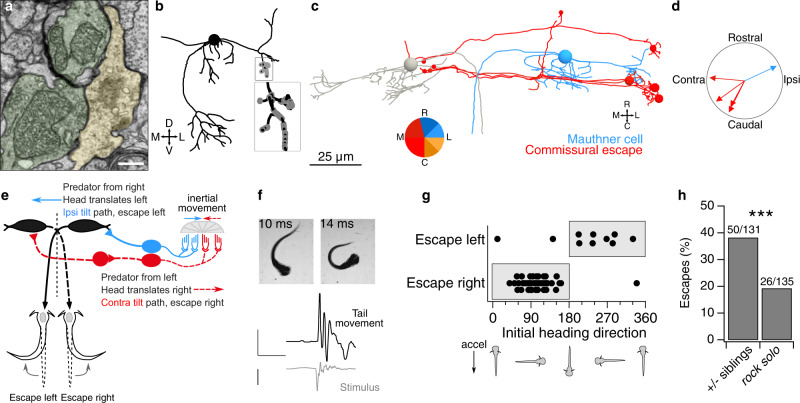
Fig. 3 Escape circuits compute head movement direction from utricular input.

a Example micrograph of utricular afferents (pseudocolored green) synapsing onto the lateral Mauthner cell dendrite (yellow). Chemical synapses are recognizable by clustered presynaptic vesicles and synaptic density. The tight apposition between the upper afferent and the Mauthner is likely an electrical synapse. Scale bar, 0.5 µm. b Coronal projection of Mauthner cell skeleton reconstruction (black, spherical soma) with utricular afferent input synapses (gray). Inset, expanded view of utricular inputs. c Horizontal projection of reconstructions of both Mauthner cells (gray, blue) and four commissural utricular neurons (red). Commissural utricular neurons make synaptic contacts on the contralateral Mauthner (small red circles). Colors indicate inferred directional tuning. d Polar plot of inferred direction tuning of utricular input to Mauthner cell and four commissural escape neurons. Directional tuning is indicated in the context of head tilt. Note that as inertial sensors, otoliths are equally sensitive to head translation in the opposite direction (e.g., ipsilateral head tilt and contralateral translation are indistinguishable). e Schematic of predicted Mauthner cell computation of head translation. A predator approaching from the right will cause a head deflection to the left. Deflection of the utricular otolith by inertia (blue arrow) would depolarize the ipsilateral tilt / contralateral translation pathway (blue; medial to the LPR). These utricular afferents excite the ipsilateral Mauthner cell, promoting an escape movement to the left. Commissural escape neurons, in contrast, will respond to rightward head movements (red hair cells and dashed lines) and are predicted to activate the contralateral Mauthner cell, promoting escapes to the right. f Example of behavioral response in a free-swimming larva subjected to rapid translation. High-speed videography captures the onset of escape and characteristic C-bend. Bottom, quantification of tail angle and translational stimulus. Scale bars, 1 rad, 100 ms, and 1 g. g Escape responses to a unidirectional translational stimulus are plotted relative to the larval heading angle at the start of the stimulus. As predicted, larvae accelerated to the right (heading direction 0–180°) escape to the right, whereas larvae accelerated to the left escape left. Escapes only occurred in the “incorrect” direction when animals were accelerated in predominantly rostral or caudal directions. h With both types of stimulus, utricle-deficient rock solo larvae escaped at approximately half the rate as their heterozygous siblings. N = 52 sibling and 55 −/− fish. Chi-squared test, p = 0.0006 (1 degree of freedom, chi-squared value = 11.65, 266 total observations).

Acknowledgments
This image is the copyrighted work of the attributed author or publisher, and ZFIN has permission only to display this image to its users. Additional permissions should be obtained from the applicable author or publisher of the image. Full text @ Nat. Commun.