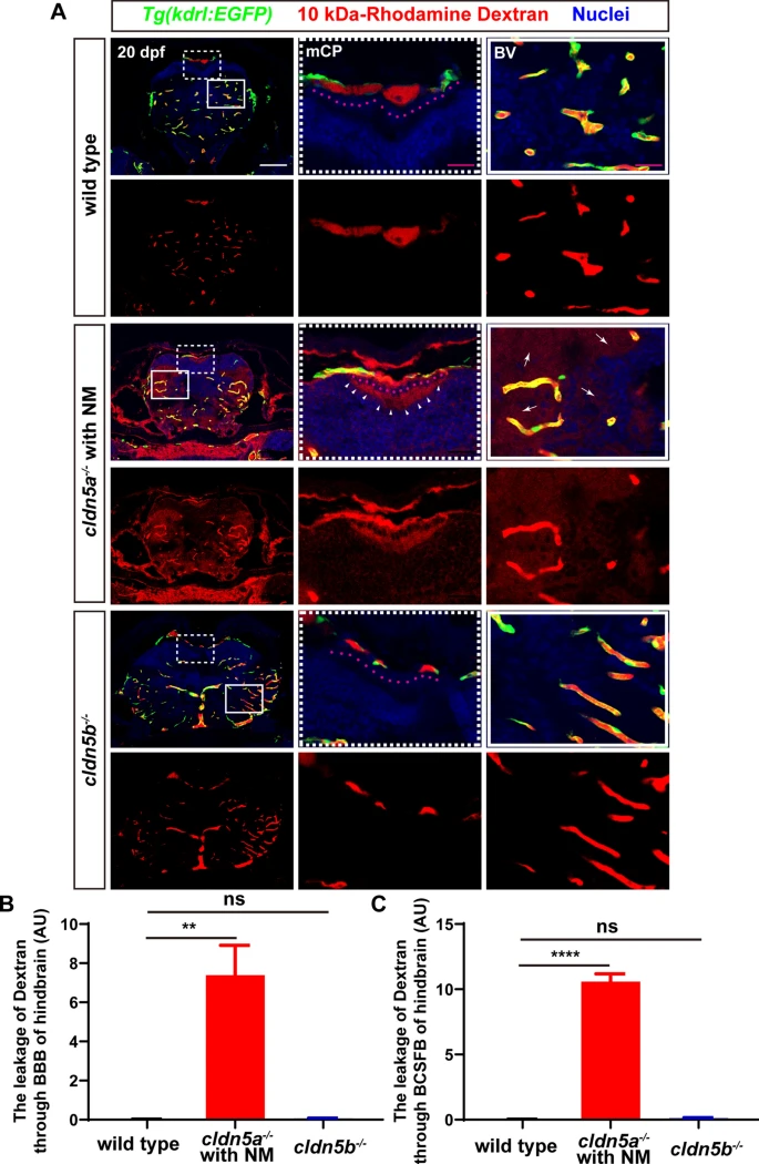
Fig. 6
The leakage of 10 kDa-rhodamine dextran through BBB and BCSFB in the hindbrain of cldn5a-/-. A In the hindbrain sections of cldn5a-/- with NM, rhodamine dextran penetrates through brain capillaries and strongly accumulated in brain parenchyma (white arrows). Meanwhile, rhodamine dextran was found to leak through the paracellular space of the mCP epithelium (pink dashline) and fullfill the hindbrain ventricle (white arrowheads). White dashed rectangles and solid rectangles indicate the enlarged regions of mCP and cerebral vessels respectively which are shown on the right side with high magnification. Pink dot lines label the position of the mCP epithelial cells. Scale bars: 100 ?m in white and 20 ?m in pink. B Quantification of the signal strength of the dye leaked through BBB into the hindbrain parenchyma. C Quantification of the signal strength of the dye leaked through BCSFB into the hindbrain ventricle. Tg(kdrl:EGFP) line was applied to label the cerebral vessels. BV, blood vessel. n?>?3 fishes analyzed per group. Data are represented as mean?±?SEM; ***P?<?0.005. ns means no significance

