
Image
|
Figure Caption
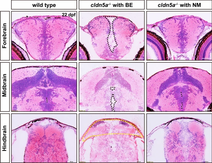
Fig. 3
Whole-brain oedema, ventricular dilatation, and cerebral hernia in cldn5a-/- with brain edema (BE). Histology of and HE staining on zebrafish brain sections at 22 dpf. In the forebrain and midbrain, cldn5a-/- with BE had a round outline and exhibited serious brain parenchymal edema and exaggerated ventricular dilatation. A yellow dot line labels the brain hernia in cldn5a-/- with BE. Black dot lines describe the brain ventricles. n?>?3 brains analyzed per group. Scale bars: 100 ?m
Acknowledgments
This image is the copyrighted work of the attributed author or publisher, and
ZFIN has permission only to display this image to its users.
Additional permissions should be obtained from the applicable author or publisher of the image.
Full text @ Fluids Barriers CNS